. 2011 Jul 26;108(30):12360-5.
doi: 10.1073/pnas.1104697108. Epub 2011 Jun 20.

Female gamete competition in an ancient angiosperm lineage

Affiliations

Female gamete competition in an ancient angiosperm lineage

Julien B Bachelier et al. Proc Natl Acad Sci U S A. .

Abstract

In Trimenia moorei, an extant member of the ancient angiosperm clade Austrobaileyales, we found a remarkable pattern of female gametophyte (egg-producing structure) development that strikingly resembles that of pollen tubes and their intrasexual competition within the maternal pollen tube transmitting tissues of most flowers. In contrast with most other flowering plants, in Trimenia, multiple female gametophytes are initiated at the base (chalazal end) of each ovule. Female gametophytes grow from their tips and compete over hundreds of micrometers to reach the apex of the nucellus and the site of fertilization. Here, the successful female gametophyte will mate with a pollen tube to produce an embryo and an endosperm. Moreover, the central tissue within the ovules of Trimenia, through which the embryo sacs grow, contains starch and other carbohydrates similar to the pollen tube transmitting tissues in the styles of most flowers. The pattern of female gametophyte development found in Trimenia is rare but by no means unique in angiosperms. Importantly, it seems that multiple female gametophytes are occasionally or frequently initiated in members of other ancient angiosperm lineages. The intensification of pollen tube (male gametophyte) competition and enhanced maternal selection among competing pollen tubes are considered to have been major contributors to the rise of angiosperms. Based on insights from Trimenia, we posit that prefertilization female gametophyte (egg) competition within individual ovules in addition to male gametophyte (sperm) competition and maternal mate choice may have been key features of the earliest angiosperms.


Conflict of interest statement

The authors declare no conflict of interest.

Figures
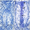
Fig. 1.
Fig. 1.
Young ovules showing the zone of megasporogenesis at the chalazal pole of the nucellus. (A) Very young ovule with one of the megaspore mother cells (mmc) in the zone of megasporogenesis (zm) at the chalazal pole (ch) of the nucellus (nuc). The asterisk marks the future site of fertilization at the micropylar end of the nucellus. (B) Ovule showing several degenerated megaspores within the zone of megasporogenesis. The arrow points to the tip of a female gametophyte (fg) that has grown out of the zone of megasporogenesis and is beginning to ascend through the nucellus central tissue (indicated by arrowheads) to the future site of fertilization (asterisk). ii, inner integument; fun, funiculus. (Scale bars: 100 μm.)
Fig. 2.
Fig. 2.
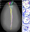
Young ovule with multiple female gametophytes. (A) 3D reconstruction (from transverse serial sections) of ovule nucellus (gray) containing tube-like female gametophytes (each a different color) initiated at the base of the nucellus and growing to the site of fertilization at the apex of the nucellus (micropylar pole of ovule at the top). One female gametophyte (green) has become dominant. (Scale bar: 200 μm.) (BD) Transverse sections from the series used for reconstruction in A and stained with periodic acid Schiff's (PAS) reagent to reveal starch and other carbohydrates in pink. Each star marks a female gametophyte of the corresponding color in the computer reconstruction; dashed lines in A indicate position of each transverse section. (B) Central tissue of the nucellus filled with starch grains (black arrows). (C) Central tissue of the nucellus with fewer and smaller starch grains and one dominant two-nucleate female gametophyte (green in A; one of two nuclei shown by the green arrowhead). (D) Base of nucellus, where additional female gametophytes (black stars) were initiated. (Scale bars: 25 μm.)
Fig. 3.
Fig. 3.
Nearly mature ovule with multiple female gametophytes. (A) 3D reconstruction of nucellus (gray) and tube-like female gametophytes (each a different color) initiated at the base of the nucellus and growing to the site of fertilization at the apex of the nucellus (micropylar pole of ovule at the top). (Scale bar: 200 μm.) (BD) Transverse sections from series used for the reconstruction in A and stained with PAS and toluidine blue. Each star marks a female gametophyte of the corresponding color in the computer reconstruction; dashed lines in A indicate the positions of each transverse section. (B) Dominant two-nucleate (green arrowheads) female gametophyte (green in A) has enlarged circumferentially after reaching depression in nucellus apex. (C) Five additional female gametophytes have grown beyond the lower one-half of nucellus (one of the two nuclei of a subordinate female gametophyte indicated by the orange arrowhead). (D) Base of nucellus, where additional female gametophytes (black stars) were initiated. (Scale bars: 25 μm.)
Fig. 4.
Fig. 4.
Mature ovule with three dominant female gametophytes. (A) 3D reconstruction of ovule nucellus (gray) and tube-like female gametophytes (each a different color) protruding through the depression in the nucellar apex (micropylar pole of ovule at the top). (Scale bar: 200 μm.) (BE) Transverse sections taken from the series used for the reconstruction in A and stained with PAS and toluidine blue. Each star marks a female gametophyte of the corresponding color in the computer reconstruction; dashed lines in A indicate position of each transverse section. (B) Enlarged apices of the three dominant female gametophytes; a pair of synergid cells (pink arrowheads) of one of two sexually mature female gametophytes (pink star). (C) A pair of synergid cells (green arrowheads) of the second sexually mature female gametophyte (green star). (D) Polar nucleus (green arrowhead) of the second mature female gametophyte (green star). (E) Polar nucleus (pink arrowhead) of the other sexually mature female gametophyte (pink star). (Scale bars: 20 μm.)
Fig. 5.
Fig. 5.
Callose in female gametophyte tubes. (A) Composite image of adjacent median longitudinal sections (white frames) of a mature ovule. Callose fluorescence is evident in cell walls of the mature female gametophyte (star) that has reached the site of fertilization (asterisk) and central tissue of nucellus. ch, chalaza; nuc, nucellus; zm, zone of megasporogenesis. (Scale bar: 200 μm.) (B) Central tissue of nucellus above zone where multiple female gametophytes are initiated. Callose fluorescence is evident in cell walls of central tissue of nucellus (n) and in the cell wall of the dominant growing female gametophyte (star). (C) Base of nucellus. Callose fluorescence can be seen in one degenerating megaspore (d) and the dominant female gametophyte (star). (Scale bars: 10 μm.)
Fig. 6.
Fig. 6.
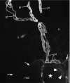
Callose in pollen tubes. Composite image of adjacent median longitudinal sections of a mature ovule. Callose fluorescence is evident in the several pollen tubes (pt) growing between the cells of the micropyle formed by the inner integument (ii). One pollen tube has reached the tip of the female gametophyte (star) and grown along its surface towards the synergids (arrowheads). Arrows indicate the surface of the nucellus (nuc). (Scale bar: 50 μm.)

Similar articles

Cited by

References

    1. Crepet WL, Niklas KJ. Darwin's second “abominable mystery”: Why are there so many angiosperm species? Am J Bot. 2009;96:366–381. - PubMed
    1. Friedman WE. The meaning of Darwin's “abominable mystery.”. Am J Bot. 2009;96:5–21. - PubMed
    1. Stebbins GL. Flowering Plants: Evolution Above the Species Level. Cambridge, MA: Harvard University Press; 1974.
    1. Mulcahy DL. The rise of the angiosperms: A genecological factor. Science. 1979;206:20–23. - PubMed
    1. Endress PK. Syncarpy and alternative modes of escaping disadvantages of apocarpy in primitive angiosperms. Taxon. 1982;31:48–52.

Publication types

MeSH terms