

Changes in lung function and chylous effusions in patients with lymphangioleiomyomatosis treated with sirolimus
- PMID: 21690594
- PMCID: PMC3176735
- DOI: 10.7326/0003-4819-154-12-201106210-00007
Changes in lung function and chylous effusions in patients with lymphangioleiomyomatosis treated with sirolimus
Abstract
Background: Lymphangioleiomyomatosis (LAM) is a disorder that affects women and is characterized by cystic lung destruction, chylous effusions, lymphangioleiomyomas, and angiomyolipomas. It is caused by proliferation of abnormal smooth muscle-like cells. Sirolimus is a mammalian target of rapamycin inhibitor that has been reported to decrease the size of neoplastic growths in animal models of tuberous sclerosis complex and to reduce the size of angiomyolipomas and stabilize lung function in humans.
Objective: To assess whether sirolimus therapy is associated with improvement in lung function and a decrease in the size of chylous effusions and lymphangioleiomyomas in patients with LAM.
Design: Observational study.
Setting: The National Institutes of Health Clinical Center.
Patients: 19 patients with rapidly progressing LAM or chylous effusions.
Intervention: Treatment with sirolimus.
Measurements: Lung function and the size of chylous effusions and lymphangioleiomyomas before and during sirolimus therapy.
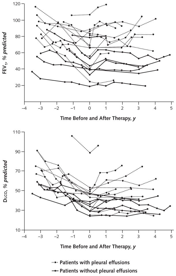
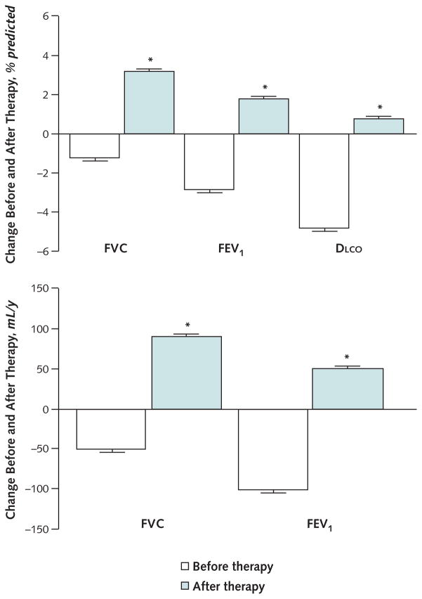
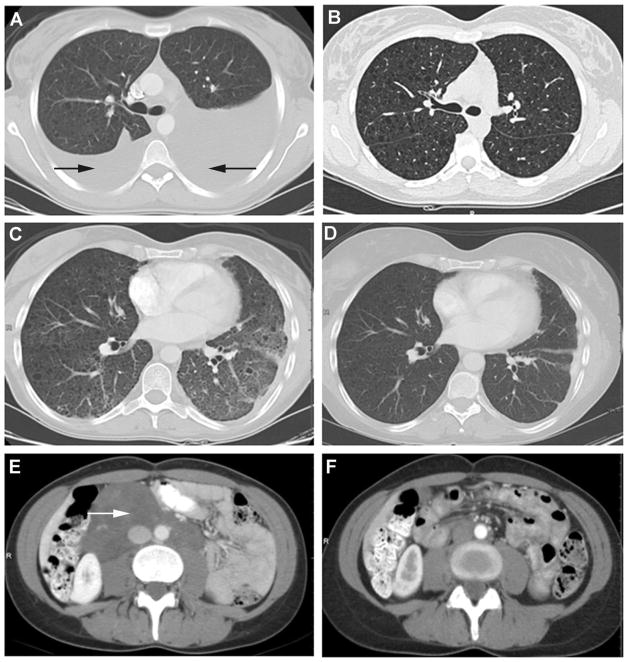
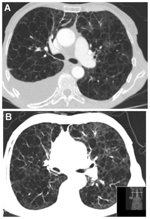
Results: Over a mean of 2.5 years before beginning sirolimus therapy, the mean (±SE) FEV1 decreased by 2.8%±0.8% predicted and diffusing capacity of the lung for carbon monoxide (Dlco) decreased by 4.8%±0.9% predicted per year. In contrast, over a mean of 2.6 years of sirolimus therapy, the mean (±SE) FEV1 increased by 1.8%±0.5% predicted and Dlco increased by 0.8%±0.5% predicted per year (P<0.001). After beginning sirolimus therapy, 12 patients with chylous effusions and 11 patients with lymphangioleiomyomas experienced almost complete resolution of these conditions. In 2 of the 12 patients, sirolimus therapy enabled discontinuation of pleural fluid drainage.
Limitations: This was an observational study. The resolution of effusions may have affected improvements in lung function.
Conclusion: Sirolimus therapy is associated with improvement or stabilization of lung function and reduction in the size of chylous effusions and lymphangioleiomyomas in patients with LAM.
Primary funding source: Intramural Research Program, National Heart, Lung, and Blood Institute, National Institutes of Health.
Conflict of interest statement
Figures
Comment in
-
Summaries for patients. Sirolimus therapy in patients with lymphangioleiomyomatosis.Ann Intern Med. 2011 Jun 21;154(12):I44. doi: 10.7326/0003-4819-154-12-201106210-00003. Ann Intern Med. 2011. PMID: 21690578 No abstract available.
-
Tuberous sclerosis complex and lymphangioleiomyomatosis: miles to go, promises to keep.Ann Intern Med. 2011 Jun 21;154(12):840-1. doi: 10.7326/0003-4819-154-12-201106210-00015. Ann Intern Med. 2011. PMID: 21690600 No abstract available.
References
-
- McCormack FX. Lymphangioleiomyomatosis: a clinical update. Chest. 2008;133:507–16. - PubMed
-
- Costello LC, Hartman TE, Ryu JH. High frequency of pulmonary lymphangioleiomyomatosis in women with tuberous sclerosis complex. Mayo Clin Proc. 2000;75:591–4. - PubMed
-
- Moss J, Avila NA, Barnes PM, Litzenberger RA, Bechtle J, Brooks PG, et al. Prevalence and clinical characteristics of lymphangioleiomyomatosis (LAM) in patients with tuberous sclerosis complex. Am J Respir Crit Care Med. 2001;164:669–71. - PubMed
-
- Franz DN, Brody A, Meyer C, Leonard J, Chuck G, Dabora S, et al. Mutational and radiographic analysis of pulmonary disease consistent with lymphangioleiomyomatosis and micronodular pneumocyte hyperplasia in women with tuberous sclerosis. Am J Respir Crit Care Med. 2001;164:661–8. - PubMed
Publication types
MeSH terms
Substances
Grants and funding
LinkOut - more resources
Full Text Sources
Other Literature Sources
Medical