. 2011;29(1):6-12.
doi: 10.1159/000324121. Epub 2011 Jun 17.

The role of cilia in the regulation of bile flow

Affiliations

The role of cilia in the regulation of bile flow

Nicholas F Larusso et al. Dig Dis. 2011.

Abstract

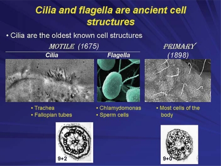
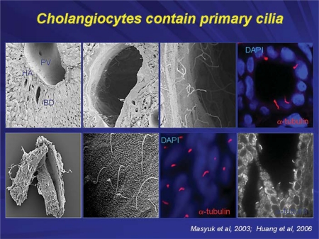
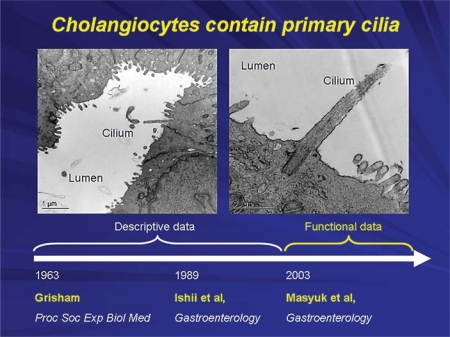
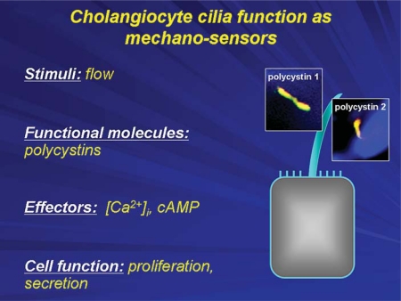
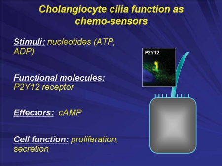
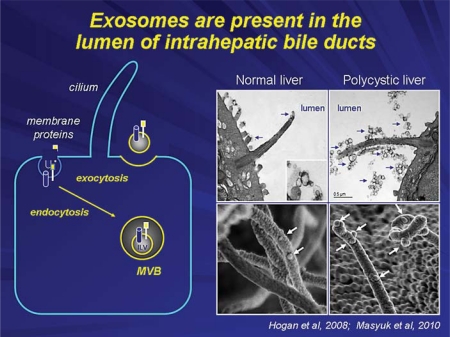
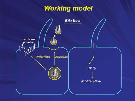
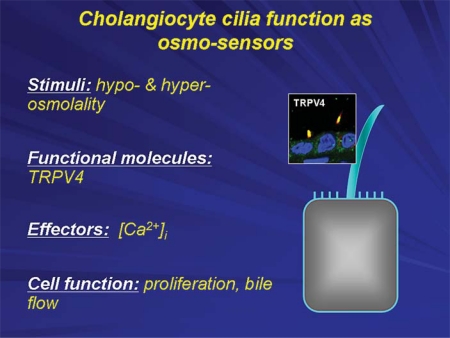
Cholangiocytes, the epithelial cells lining intrahepatic bile ducts, are ciliated cells. Each cholangiocyte has a primary cilium consisting of (i) a microtubule-based axoneme and (ii) the basal body, centriole-derived, microtubule-organizing center from which the axoneme emerges. Primary cilia in cholangiocytes were described decades ago, but their physiological and pathophysiological significance remained unclear until recently. We now recognize that cholangiocyte cilia extend from the apical plasma membrane into the bile duct lumen and, as such, are ideally positioned to detect changes in bile flow, bile composition and bile osmolality. These sensory organelles act as cellular antennae that can detect and transmit signals that influence cholangiocyte function. Indeed, recent data show that cholangiocyte primary cilia can activate intracellular signaling pathways when they sense modifications in the flow, molecular constituents and osmolarity of bile. Their ability to sense and transmit signals depends on the participation of a growing number of specific ciliary-associated proteins that act as receptors, channels and transporters. Cholangiocyte cilia, in addition to being important in normal biliary physiology, likely contribute to the cholangiopathies when their normal structure or function is disturbed. Indeed, the polycystic liver diseases that occur in combination with autosomal dominant and recessive polycystic kidney disease (i.e. ADPKD and ARPKD) are two important examples of such conditions. Recent insights into the role of cholangiocyte cilia in cystic liver disease using in vitro and animal models have already resulted in clinical trials that have influenced the management of cystic liver disease.


Figures

Fig. 1
Fig. 1
Cilia are present in many organisms from Chlamydomonas reinhardi to mammals. They are centriole-derived organelles consisting of a set of microtubules surrounded by a membrane. Based on the microtubule arrangements, cilia are classified as 9+2 (9 peripheral microtubule doublets arranged around a central core that contains 2 central microtubules) and 9+0 (contain only 9 peripheral microtubule doublets). Multiciliated cells usually possess motile 9+2 cilia, while nonmotile 9+0 (i.e. primary) cilia are present in uniciliated cells. Mammalian motile cilia are present in large numbers on cell surfaces, such as epithelial cells lining airways, ependyma and choroid plexus in the brain, oviduct and epididymis of the reproductive tracts. These cilia beat in an orchestrated wavelike fashion and are involved in movement of mucus in the lung and cerebrospinal fluid in the brain, or in transport of ovum and sperm along the reproductive tracts. The primary cilium is a solitary, nonmotile, long, tubular organelle extending from the plasma membrane of the cell. With few exceptions (e.g. nucleated blood cells, hepatocytes), primary cilia are ubiquitous in vertebrates. They are found on epithelial cells of the bile ducts, kidney tubules, the pancreas and the thyroid glands as well as on nonepithelial cells such as chondrocytes, fibroblasts, smooth muscle cells and neurons.
Fig. 2
Fig. 2
In whole rat liver, no visible cilia are observed in hepatocytes, in the lumen of the portal vein or hepatic artery; primary cilia (approx. 7 μm in length) were present only in the intrahepatic bile ducts extending into the ductal lumen from the cholangiocyte apical plasma membrane (top panels: scanning and confocal microscopy). In intrahepatic bile ducts isolated from normal rats by microdissection, a single primary cilium is clearly observed on the cholangiocyte apical membrane by scanning electron, confocal and transmission immunofluorescent microscopy (bottom panels). Cilia on confocal images (red) are stained with ciliary marker, acetylated α-tubulin; nuclei are stained blue with DAPI [17,18].
Fig. 3
Fig. 3
Primary cilia in cholangiocytes were first described in 1963. During our work on biliary epithelia, we often observed cholangiocyte cilia under different occasions, but did not pay much attention to these organelles. Our laboratory published the first report describing the presence of cilia in cholangiocytes in 1989. In 2003, we showed that they are critically important for normal cholangiocyte functions and that their abnormalities lead to formation of hepatic cysts [6,7,17].
Fig. 4
Fig. 4
Cholangiocyte primary cilia function as mechanosensors. The bending of cholangiocyte cilia by luminal fluid flow induces an increase in [Ca2+]i, which depends on polycystin-1 and polycystin-2 and intracellular Ca2+ sources. The mechanosensory functions of cholangiocyte cilia also occur through cAMP signaling affecting cell proliferation and fluid secretion.
Fig. 5
Fig. 5
Cholangiocyte primary cilia function as chemosensors. Chemosensation by cholangiocyte cilia occurs with the involvement of P2Y12, a receptor that is activated by biliary nucleotides (i.e. ATP and ADP) causing changes in intracellular cAMP levels that subsequently affects cell proliferation and fluid secretion.
Fig. 6
Fig. 6
Exosomes are involved in chemosensory function of cholangiocyte cilia. Exosomes are small (30–100 nm in diameter) extracellular membrane-enclosed vesicles. They are derived from the internal vesicles of multivesicular bodies (MVB) that fuse with the plasma membrane in an exocytic manner and release their content into the extracellular space (scheme). The presence of exosome-like vesicles of 50–80 nm in diameter in the lumen of intrahepatic bile ducts of wild-type and polycystic mice was confirmed by transmission electron microscopy (right, top panels). These vesicles surround cholangiocyte cilia and some appear to attach to the ciliary membrane and microvilli. The SEM images (right, bottom panels) suggest that exosome-like vesicles in fact attach to cilia [10,11].
Fig. 7
Fig. 7
Exosomes are involved in intercellular communication within the liver. They deliver functional proteins, lipids and nucleic acids to neighboring or distant cells, interact with cholangiocyte cilia subsequently triggering downstream signaling events in targeted cells and functional responses.
Fig. 8
Fig. 8
Cholangiocyte primary cilia function as osmosensors. A mammalian transient receptor potential vanilloid 4 (TRPV4) channel exquisitely sensitive to minute changes in osmolality of extracellular milieu, being activated by extracellular hypotonicity and inhibited by extracellular hypertonicity. Exposure of cholangiocytes to hypotonicity results in an increase in intracellular Ca2+, which depends on extracellular Ca2+ sources. Changes in luminal tonicity also induce TRPV4- and ciliary-dependent bicarbonate secretion, suggesting an important role of osmosensory function of cholangiocyte cilia in ductal bile formation.

References

    1. Christensen ST, Pedersen LB, Schneider L, Satir P. Sensory cilia and integration of signal transduction in human health and disease. Traffic. 2007;8:97–109. - PubMed
    1. Dawe HR, Farr H, Gull K. Centriole/basal body morphogenesis and migration during ciliogenesis in animal cells. J Cell Sci. 2007;120:7–15. - PubMed
    1. Satir P, Christensen ST. Overview of structure and function of mammalian cilia. Annu Rev Physiol. 2007;69:377–400. - PubMed
    1. Masyuk AI, Masyuk TV, LaRusso NF. Cholangiocyte primary cilia in liver health and disease. Dev Dyn. 2008;237:2007–2012. - PMC - PubMed
    1. Marshall WF, Nonaka S. Cilia: tuning in to the cell's antenna. Curr Biol. 2006;16:R604–R614. - PubMed

Publication types