. 2011 Aug;115(2):242-53.
doi: 10.1097/ALN.0b013e318223b8b9.

Phosphorylation of GSK-3β mediates intralipid-induced cardioprotection against ischemia/reperfusion injury

Affiliations

Phosphorylation of GSK-3β mediates intralipid-induced cardioprotection against ischemia/reperfusion injury

Siamak Rahman et al. Anesthesiology. 2011 Aug.

Abstract

Background: Intralipid (Sigma, St. Louis, MO), a brand name for the first safe fat emulsion for human use, has been shown to be cardioprotective. However, the mechanism of this protection is not known. The authors investigated the molecular mechanism(s) of Intralipid-induced cardioprotection against ischemia/reperfusion injury, particularly the role of glycogen synthase kinase-3β (GSK-3β) and mitochondrial permeability transition pore in this protective action.

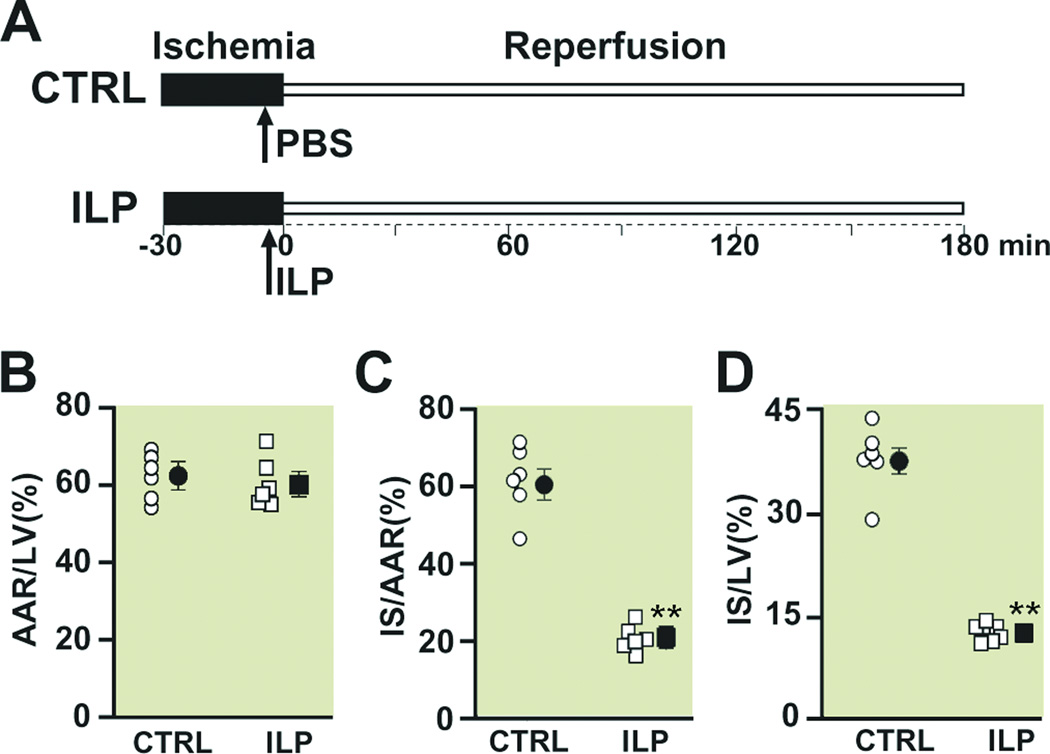
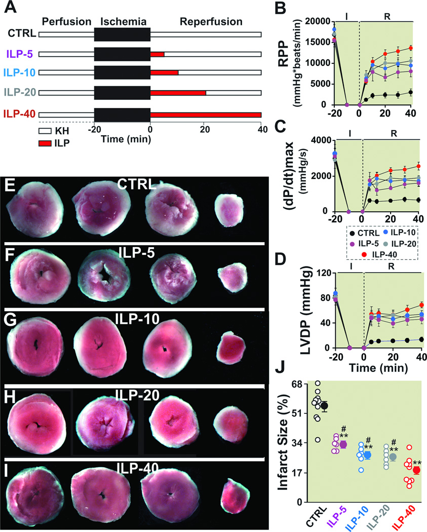
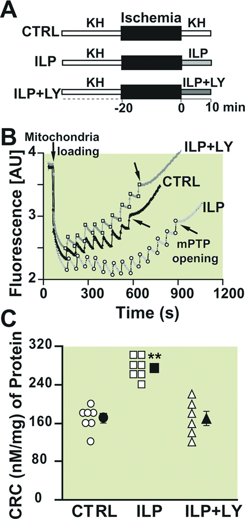
Methods: In vivo rat hearts or isolated Langendorff-perfused mouse hearts were subjected to ischemia followed by reperfusion with Intralipid (1% in ex vivo and one bolus of 20% in in vivo) or vehicle. The hemodynamic function, infarct size, threshold for the opening of mitochondrial permeability transition pore, and phosphorylation levels of protein kinase B (Akt)/extracellular signal regulating kinase (ERK)/GSK-3β were measured.

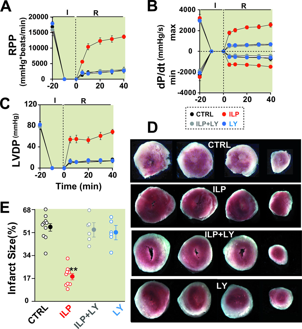
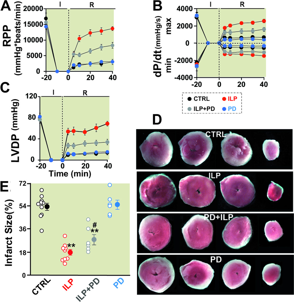
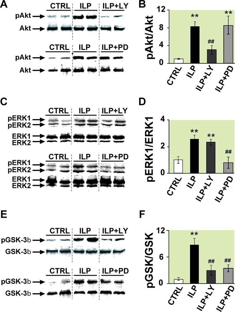
Results: Administration of Intralipid at the onset of reperfusion resulted in approximately 70% reduction in infarct size in the in vivo rat model. Intralipid also significantly improved functional recovery of isolated Langendorff-perfused mouse hearts as the rate pressure product was increased from 2,999 ± 863 mmHg*beats/min in the control group to 13,676 ± 611 mmHg*beats/min (mean±SEM) and the infarct size was markedly smaller (18.3 ± 2.4% vs. 54.8 ± 2.9% in the control group, P < 0.01). The Intralipid-induced cardioprotection was fully abolished by LY294002, a specific inhibitor of PI3K, but only partially by PD98059, a specific ERK inhibitor. Intralipid also increased the phosphorylation levels of Akt/ERK1/glycogen synthase kinase-3β by eightfold, threefold, and ninefold, respectively. The opening of mitochondrial permeability transition pore was inhibited by Intralipid because calcium retention capacity was higher in the Intralipid group (274.3 ± 8.4 nM/mg vs. 168.6 ± 9.6 nM/mg in the control group).

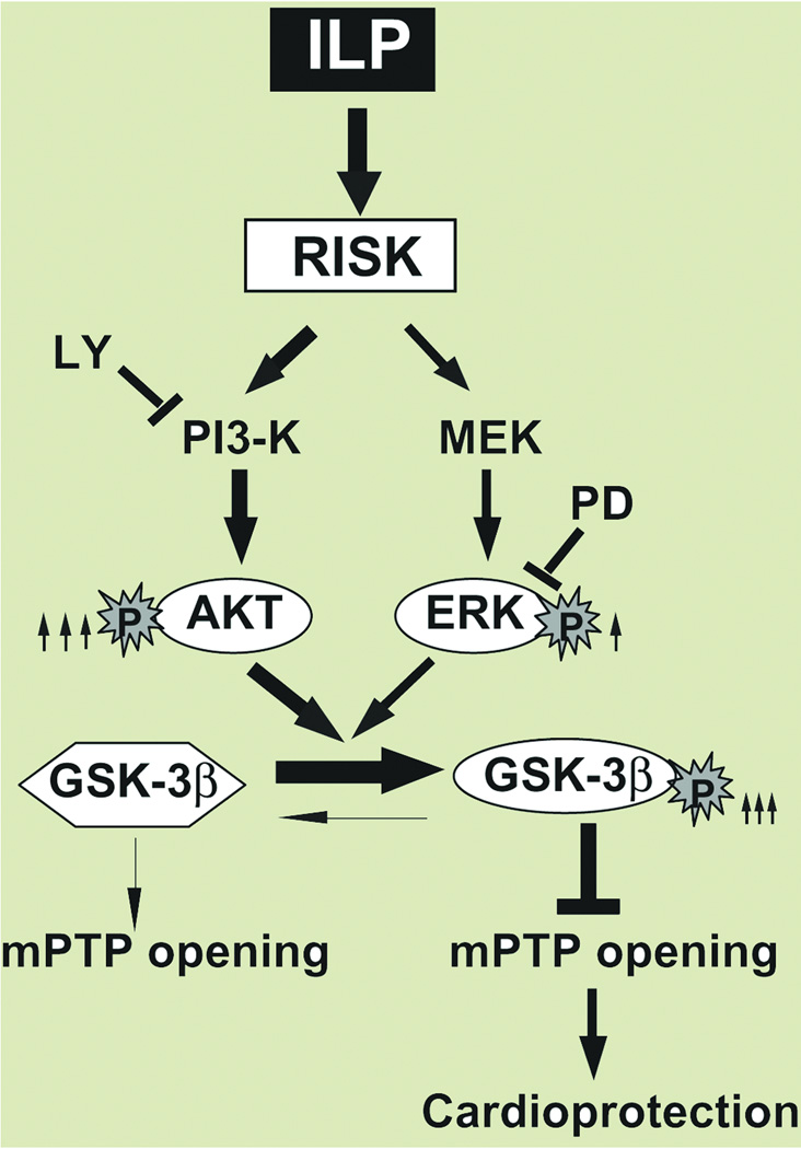
Conclusions: Postischemic treatment with Intralipid inhibits the opening of mitochondiral permeability transition pore and protects the heart through glycogen synthase kinase-3β via PI3K/Akt/ERK pathways.


Conflict of interest statement

Conflict of Interest: None

Figures

Figure 1
Figure 1. Intralipid administration reduces the infarct size in the in vivo ischemia/reperfusion rat model
A. experimental protocol, the left coronary artery was occluded for 30 minutes followed by 3 hr of reperfusion. One single IV bolus of Phosphate Buffered Saline (PBS) (control group, CTRL) or 20% Intralipid (5ml/kg body weight, ILP) was administered 5 min before reperfusion. Percentage of area at risk (AAR) divided by left ventricle (LV) (B), infarct size (IS) divided by AAR (C), and infarct size (IS) divided by left ventricle in CTRL (circles) and Intralipid (squares). The individual measurements (n=6 in ILP and n=6 in CTRL) are shown in open shapes whereas the averages (Mean±SEM) are shown in filled shapes **P<0.01 vs. CTRL.
Figure 2
Figure 2. Administration of Intralipid at reperfusion improves heart functional recovery and reduces infarct size against reperfusion injury
A. Experimental protocol. The isolated mouse hearts were reperfused with Krebs Henseleit (KH, control group, CTRL), or 1% Intralipid for 5 min (ILP-5), 10 min (ILP-10), 20 min (ILP-20) or 40 min (ILP-40), followed by reperfusion with KH for the remainder of 40 min. Rate pressure product (RPP, B), the maximum rate of left ventricle (LV) pressure rise (dP/dtmax) and decline (−dP/dtmin, C) and left ventricular developed pressure (LVDP, D) as a function of time in CTRL (black, n=6), ILP-5 (purple, n=6), ILP-10 (blue, n=6), ILP-20 (gray, n=6) and ILP-40 (red, n=6). Four slices of the same heart after 2,3,5-triphenyltetrazolium chloride (TTC) staining in CTRL (E), ILP-5 (F), ILP-10 (G), ILP-20 (H), and ILP-40 (I). The white area represents the infarct zone and the red shows the viable area. J. The area of necrosis as the percentage of total left ventricular (LV) area in CTRL (black, n=10), ILP-5 (purple, n=6), ILP-10 (blue, n=6), ILP-20 (gray, n=6) and ILP-40 (red, n=9). The individual measurements in each groups are shown in open circles whereas the averages (Mean±SEM) are shown in filled circles **P < 0.01 vs. CTRL, #P<0.05 vs. ILP-40.
Figure 3
Figure 3. PI3K inhibitor abolishes Intralipid-induced cardioprotection against ischemia/ reperfusion injury
Rate pressure product (RPP, A), the maximum rate of left ventricle (LV) pressure rise (dP/dtmax) and decline (−dP/dtmin, B) and left ventricular developed pressure (LVDP, C) as a function of time in control group (CTRL, black, n=6), 1% Intralipid (ILP, red, n=6), 1% Intralipid+LY294002 (ILP+LY, gray, n=6) and LY294002 alone (LY, blue, n=6). D. Four slices of the same heart in CTRL, ILP, ILP+LY and LY alone after 2,3,5-triphenyltetrazolium chloride (TTC) staining. The white area represents the infarct zone and the red shows the viable area. E. The area of necrosis as the percentage of total left ventricular (LV) area in CTRL (black, n=10), ILP (red, n=9), ILP+LY (gray, n=6) and in LY (blue, n=6). The individual measurements in each group are shown in open circles whereas the averages (Mean±SEM) are shown in filled circles **P<0.01 vs. CTRL.
Figure 4
Figure 4. ERK inhibitor abolishes partially Intralipid-induced cardioprotection against ischemia/ reperfusion injury
Rate pressure product (RPP, A), the maximum rate of LV pressure rise (dP/dtmax) and decline (−dP/dtmin, B) and left ventricular developed pressure (LVDP, C) as a function of time in control group (CTRL, black, n=6), 1% Intralipid (ILP, red, n=6), 1% Intralipid+PD98059 (ILP+PD, gray, n=6) and PD98059 alone (PD, blue, n=6). D. Four slices of the same heart in CTRL, ILP, ILP+PD and PD after TTC staining. The white area represents the infarct zone and the red shows the viable area. E. The area of necrosis as the percentage of total left ventricular (LV) area in CTRL (black, n=10), ILP (red, n=9), ILP+PD (gray, n=6)) and in PD (blue, n=6)). The individual measurements in each group are shown in open circles whereas the averages (Mean±SEM) are shown in filled circles. **P < 0.01 vs. CTRL and #P<0.05 vs. ILP).
Figure 5
Figure 5. Involvement of PI3K-Akt and ERK pathways and their downstream target GSK-3β in Intralipid -induced protection
A, C, E. representative immunoblots of pAkt and total Akt (A), pERK1,2 and total ERK1,2 (C), and pGSK-3β and total GSK-3β (E) in heart homogenates subjected to ischemia/reperfusion from control group (CTRL), Intralipid group (ILP) and 1% Intralipid+LY294002 (ILP+LY) top panels or CTRL, ILP and 1% Intralipid+PD98059 (ILP+PD) lower panels. B, D, F. Western Blot quantification (Mean±SEM) of pAkt protein to total Akt (B), pERK1 to total ERK (D) and pGSK-3β to total GSK-3β (E) ratios in CTRL (white bars, n=6), ILP (black bars, n=4), ILP+LY (dark gray bars, n=4) and ILP+PD (light gray bars, n=4). **P<0.01 vs. CTRL; ##P<0.01 vs. ILP.
Figure 6
Figure 6. Intralipid inhibits the opening of the mitochondrial permeability transition pore and this inhibition is abolished by PI3K inhibitor
A. Experimental protocol for measuring calcium retention capacity. At the onset of reperfusion, isolated hearts are perfused with Krebs Henseleit (KH, control group, CTRL), 1% Intarlipid (ILP) or 1% Intralipid+LY294002 (ILP+LY) for 10 min. B. Typical recording of the mitochondria permeability transition pore opening in isolated mitochondria from control (black trace), ILP (light gray trace) and ILP+LY (dark gray trace). Fourteen pulses (gray arrows) of 20 nM calcium were required to trigger the opening of mitochondria permeability transition pore in ILP group compared to 7 pulses (black arrows) in CTRL and 8 pulses in ILP+LY. C. Calcium retention capacity (CRC) in CTRL (circles, n=7), ILP (squares, n=7) and ILP+LY (triangles, n=6). The individual measurements in each groups are shown in open shapes whereas the averages (Mean±SEM) are shown in filled shapes.**P<0.01 vs. CTRL and ILP+LY.
Figure 7
Figure 7. Proposed mechanisms underlying Intralipid-induced cardioprotection against ischemia/reperfusion injury
The Reperfusion Injury Salvage Kinases (RISK) pathway is activated in the presence of Intralipid (ILP), resulting in increased phosphorylation of both Akt and ERK, although the degree of activation is much more prounnced in Akt (8 fold, shown by thick arrow) than in ERK (3 fold, shown by thin arrow). Both pathways converge to phosphorylate GSK-3β (inactive form), which in turn inhibits the opening of the mPTP and induces protection against reperfusion injury. The protection provided by Intralipid is fully abolished by the PI3K specific inhibitor, LY294002 (LY) and partially by PD98059 (PD).

Comment in

References

    1. Christian TF, Schwartz RS, Gibbons RJ. Determinants of infarct size in reperfusion therapy for acute myocardial infarction. Circulation. 1992;86:81–90. - PubMed
    1. Zhao ZQ, Corvera JS, Halkos ME, Kerendi F, Wang NP, Guyton RA, Vinten-Johansen J. Inhibition of myocardial injury by ischemic postconditioning during reperfusion: Comparison with ischemic preconditioning. Am.J.Physiol Heart Circ.Physiol. 2003;285:H579–H588. - PubMed
    1. Bopassa JC, Ferrera R, Gateau-Roesch O, Couture-Lepetit E, Ovize M. PI 3-kinase regulates the mitochondrial transition pore in controlled reperfusion and postconditioning. Cardiovas Res. 2006;69:178–185. - PubMed
    1. Cohen MV, Yang XM, Downey JM. The pH hypothesis of postconditioning: Staccato reperfusion reintroduces oxygen and perpetuates myocardial acidosis. Circulation. 2007;115:1895–1903. - PubMed
    1. Tamareille S, Ghaboura N, Treguer F, Khachman D, Croue A, Henrion D, Furber A, Prunier F. Myocardial reperfusion injury management: Erythropoietin compared with postconditioning. Am.J.Physiol Heart Circ.Physiol. 2009;297:H2035–H2043. - PubMed

Publication types

MeSH terms