Review
. 2011 Jun 21;8(8):428-39.
doi: 10.1038/nrurol.2011.85.

Epithelial-to-mesenchymal transition in prostate cancer: paradigm or puzzle?

Affiliations
Review

Epithelial-to-mesenchymal transition in prostate cancer: paradigm or puzzle?

Jones T Nauseef et al. Nat Rev Urol. .

Abstract

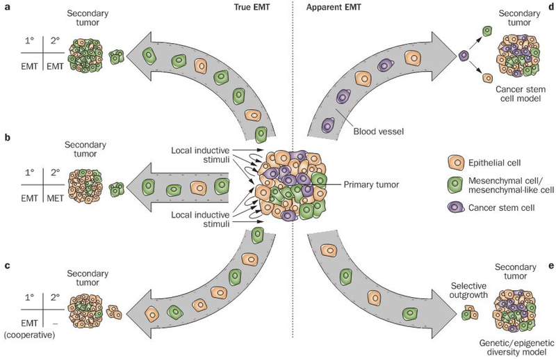
The lethal consequences of prostate cancer are related to its metastasis to other organ sites. Epithelial-to-mesenchymal transition (EMT) has received considerable attention as a conceptual paradigm to explain invasive and metastatic behavior during cancer progression. EMT is a normal physiologic process by which cells of epithelial origin convert into cells bearing mesenchymal characteristics. It has been proposed that EMT is co-opted by cancer cells during their metastatic dissemination from a primary organ to secondary sites, but the extent to which this recapitulates physiologic EMT remains uncertain. However, there is ample evidence that EMT-like states occur in, and may contribute to, prostate cancer progression and metastasis, and so has become a very active area of research. Here we review this evidence and explore recent studies that have aimed to better define the role and mechanisms of EMT in prostate cancer. While definitive evidence of something akin to physiologic EMT is still lacking in human prostate cancer, this area of research has nonetheless provided new avenues of investigation into the longstanding puzzles of metastasis, therapeutic resistance, and prognostic biomarkers.


Conflict of interest statement

Competing interests

The authors declare no competing interests.

Figures

Figure 1 |
Figure 1 |
True and apparent EMT might both contribute to metastasis, yet be difficult to distinguish. Primary tumors contain a heterogeneous mixture of cells, including epithelial cells and cells that resemble mesenchymal cells. Whether this latter group of cells is the result of the transition of an individual cell from an epithelial to a mesenchymal phenotype, however, is unclear. The patterns observed in the primary and metastatic tumors can be reconciled by a variety of epistemological explanations. a | EMT occurs in the primary tumor, resulting in the entrance of both mesenchymal and epithelial cells into circulation. The mesenchymal cells are those that are chiefly capable of extravasation and colonization at a secondary site. b | Similarly, EMT occurs at the primary tumor, and mesenchymal cells exit the bloodstream at a new site to initiate a secondary tumor; however, in this model, the mesenchymal cells at the site of metastasis undergo MET back to an epithelial phenotype. c | in this instance, EMT again occurs at the primary site, but to a different end. Here, the mesenchymal cells facilitate the metastatic spread of epithelial cells. Because the mesenchymal cells serve a cooperative role at the point of departure, it is the epithelial cells that disseminate most effectively. Consequently, there is no need for MET at the secondary site in this model. d | The cancer stem cell population expands at the site of the primary tumor, perhaps under the influence of EMT, and make up the important subset of cells in circulatory transit. Upon arrival at the secondary site, these cells exercise stem-cell-like characteristics of asymmetrical division and pluripotency to generate both epithelial and mesenchymal cells. e | A subpopulation of epithelial cells begins to resemble mesenchymal cells due to genetic and/or epigenetic changes. conferring some of the invasive behaviors of mesenchymal cells and facilitating the steps of metastasis (Figure 3). The presence of this population reflects selective outgrowth, rather than transdifferentiation of individual cells. Abbreviations: EMT, epithelial-to-mesenchymal transition; MET, mesenchymal-to-epithelial transition.
Figure 2 |
Figure 2 |
Many roads lead to EMT: the molecular basis for the mechanisms of EMT and EMT-like states. Inductive stimuli in the tumor microenvironment, including hypoxia and growth factors (e.g. TGF-β, FGF and IGF-1), trigger downstream signaling. These pathways, via MAPK, Smad, GSK3β, and NFκB, result in increased activity of transcriptional repressors in the ZEB, Twist, and Snail families, which repress E-cadherin and other epithelial cell adhesion proteins and induce other mesenchymal proteins. Epigenetic mechanisms involving EZH2 can promote EMT-like states, whereas miRNAs (such as miR-101 or those in the miR-200 family) might act to maintain epithelial status. Importantly, this picture presents numerous mutually reinforcing mechanisms that might promote the EMT-like state. Abbreviations: CDH1, cadherin-1 (E-cadherin); ERβ, estrogen receptor β; FGF, fibroblast growth factor; GSK3β, glycogen synthase kinase 3β; HIF-1α, hypoxia inducible factor 1α; IGF, insulin-like growth factor; ITGβ4; integrin β4; LAM332, laminin-332; MAPK, mitogen-activated protein kinase; miRNA, micro RNA; NFκB, nuclear factor κB; PKD1, protein kinase D1; RKIP, Raf kinase inhibitor protein; sFRP, secreted frizzled-related protein; TGF-β, transforming growth factor β; VEGF-A, vascular endothelial growth factor A; WIF, Wnt inhibitory factor.
Figure 3 |
Figure 3 |
The possible roles of EMT-like states in the metastatic cascade. The loss of cell–cell adhesion that accompanies the loss of E-cadherin-mediated adherens junctions is a characteristic feature of EMT induced in vitro. This decreased cell–cell adhesion is accompanied by changes in the expression of proteases and matrix proteins that allow motile cells to invade through the basement membrane, resulting in invasive carcinoma. Similarly, these events can facilitate entrance into and escape from the bloodstream. Once at a secondary tissue, metastatic colonization requires survival, proliferation, and, possibly, MET. Abbreviations: EMT, epithelial-to-mesenchymal transition; MET, mesenchymal-to-epithelial transition.

References

    1. Thiery JP, Acloque H, Huang RY & Nieto MA Epithelial-mesenchymal transitions in development and disease. Cell 1139 871–890 (2009). - PubMed
    1. Hay ED Role of cell-matrix contacts in cell migration and epithelial-mesenchymal transformation. Cell Differ. Dev 32, 367–375 (1990). - PubMed
    1. Bussemakers MJ et al. Decreased expression of E-cadherin in the progression of rat prostatic cancer. cancer Res 52, 2916–2922 (1992). - PubMed
    1. Thompson EW, Newgreen DF & Tarin D Carcinoma invasion and metastasis: a role for epithelial-mesenchymal transition? Cancer Res 65, 5991–5995 (2005). - PubMed
    1. Tarin D, Thompson EW & Newgreen DF fallacy of epithelial mesenchymal transition in neoplasia. cancer Res 65, 599–000 (2005). - PubMed

Publication types