Review
. 2011 Jun 21;8(8):443-55.
doi: 10.1038/nrcardio.2011.87.

Mechanisms of disease: pulmonary arterial hypertension

Affiliations
Review

Mechanisms of disease: pulmonary arterial hypertension

Ralph T Schermuly et al. Nat Rev Cardiol. .

Abstract

Our understanding of, and approach to, pulmonary arterial hypertension has undergone a paradigm shift in the past decade. Once a condition thought to be dominated by increased vasoconstrictor tone and thrombosis, pulmonary arterial hypertension is now seen as a vasculopathy in which structural changes driven by excessive vascular cell growth and inflammation, with recruitment and infiltration of circulating cells, play a major role. Perturbations of a number of molecular mechanisms have been described, including pathways involving growth factors, cytokines, metabolic signaling, elastases, and proteases, that may underlie the pathogenesis of the disease. Elucidating their contribution to the pathophysiology of pulmonary arterial hypertension could offer new drug targets. The role of progenitor cells in vascular repair is also under active investigation. The right ventricular response to increased pressure load is recognized as critical to survival and the molecular mechanisms involved are attracting increasing interest. The challenge now is to integrate this new knowledge and explore how it can be used to categorize patients by molecular phenotype and tailor treatment more effectively.


Conflict of interest statement

R. T. Schermuly declares he has been a consultant for Actelion, Bayer Healthcare, Ergonex, and Solvay Pharmaceuticals, and has received speakers bureau (honoraria) from Actelion, Bayer Healthcare, GlaxoSmithKlein, Lilly, Solvay Pharmaceuticals, and Pfizer. R. T. Schermuly also declares he has received grant/research support from Actelion, Bayer Healthcare, Ergonex, Excellence Cluster Cardiopulmonary System, German Research Foundation, Gilead, Novartis, Solvay Pharmaceuticals, Pfizer, and the University of Giessen and Marburg Lung Center.

H. A. Ghofrani declares he has been a consultant for Bayer Healthcare, Ergonex, GlaxoSmithKlein, Novartis, and Pfizer, and has received speakers bureau (honoraria) from Bayer Healthcare, GlaxoSmithKlein, Novartis, and Pfizer. H. A. Ghofrani also declares he has received grant/research support from Bayer Healthcare, Excellence Cluster Cardiopulmonary System, German Research Foundation, Novartis, Pfizer, and the University of Giessen and Marburg Lung Center.

M. R. Wilkins declares he has been a consultant for Biomarin and Pfizer, has received speakers bureau (honoraria) from Bayer Healthcare, GlaxoSmithKlein, and Pfizer, and has received grant/research support from Biomarin.

F. Grimminger declares he has been a consultant for Bayer Healthcare, has received speakers bureau (honoraria) from Actelion, Encysive, and Pfizer, and has received grant/research support from Actelion, Excellence Cluster Cardiopulmonary System, GlaxoSmithKlein, German Research Foundation, Novartis, Parexel International GmbH, Pfizer, and the University of Giessen and Marburg Lung Center.

Figures

Figure 1
Figure 1. Vascular remodeling in pulmonary arterial hypertension.
Putative therapeutic targets are indicated. Abbreviations: 5-HT, 5-hydroxytryptamin; K- and Ca-channels, potassium and calcium channels; AEC, alveolar epithelial cells; BMP, bone morphogenetic protein; cGMP, cyclic guanosine monophosphate; ECM, extracellular matrix; EGF, epidermal growth factor; EPC, endothelial progenitor cells; HIF, hypoxia inducible factor; MMPs, matrix metalloproteinases; NADPH, nicotinamide adenine dinucleotide phosphate; NO, nitric oxide; PDE, phosphodiesterase; PDGF, platelet-derived growth factor; PGI2, prostaglandin I2; Rho-Ki, Rho kinases; ROS, reactive oxygen species; sGC, soluble guanylate cyclase; TGF, transforming growth factor-β; TK, tyrosine kinase; TKi, tyrosine kinase inhibitor; TRPC, transient receptor potential cation channels; VEGF, vascular endothelial growth factor.
Figure 2
Figure 2. Growth factor signaling in pulmonary arterial hypertension.
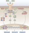
Expression of certain growth factors is increased in pulmonary arterial hypertension, promoting cell proliferation, survival, and migration via a number of signaling pathways, and thus contributing to pulmonary vascular remodeling. Abbreviations: Akt, v-akt murine thymoma viral oncogene homolog; AP-1, activator protein-1; CaM, calmodulin; CamKII, calcium/calmodulin-dependent kinase II; CRE, cyclic adenosine monophosphate response element; DAG, diacylglycerol; Erk 1/2, extracellular signal-regulated kinase 1/2; FGF, fibroblast growth factor; GF, growth factor; Grb2, growth factor receptor-bound protein 2; IP3, inositol triphosphate; JAK-2, Janus-activated kinase 2; KSR, kinase suppressor of Ras; Mek 1/2, mitogen-activated protein kinase kinase 1/2; MLCK, myosin light chain kinase; PDGF, platelet-derived growth factor; PI3K, phosphoinositide-3-kinase; PKB, protein kinase B; PKC, protein kinase C; PLC, phospholipase C; Raf, rapidly growing fibrosarcoma; Ras, Ras protein; SOCS, suppressor of cytokine signaling; SOS, son of sevenless; STAT, signal transducer and activator of transcription.
Figure 3
Figure 3. Notch signaling pathway and its proposed role in pulmonary hypertension.
In the absence of activated Notch signaling, the DNA binding protein CBF-1/RBP-Jκ forms a complex with co-repressors to prevent transcription of target genes. Interaction of ligand (via its DSL motif) with the EGF-like repeats on the receptor triggers two successive proteolytic cleavages by TACE, followed by gamma secretase. These cleavages result in release of NICD, which translocates to the nucleus and interacts with CBF-1/RBP-Jκ, removing the co-repressors. Coactivators, including MAML, CBP, and other transcription factors, are subsequently recruited, leading to transcription of Notch target genes (the HES family of transcription repressors). Notch 3 is the predominantly expressed Notch receptor in vascular SMCs, including pulmonary artery SMCs. Arrows indicate changes observed in pulmonary arterial hypertension. Abbreviations: APH 1, anterior pharynx-defective 1; CBF-1, Cp-binding factor 1; CBP, CREB binding protein; CREB, cyclic adenosine monophosphate response element-binding; DSL, Delta Serrate Ligand; EGF, epidermal growth factor; MAML, mastermind-like protein; NICD, Notch Intracellular Domain; PEN 2, presenilin 2; RBP-Jκ, recombination signal sequence-binding protein-Jκ; SMC, smooth muscle cell; TACE, tumor necrosis factor α-converting enzyme; TBP, TATA box-binding protein; TF, transcription factor.
Figure 4
Figure 4. Inflammation-mediated vascular remodeling in PAH.
In response to infection and inflammatory events, lung vascular cells produce inflammatory mediators (chemokines and cytokines), thereby recruiting inflammatory cells (macrophages, dendritic cells, mast cells, B cells, T cells, and T regulatory cells). With the coordination of inflammatory mediators, inflammatory cells might perpetuate the release of cytokines, chemokines, and growth factors. Finally, these processes lead to vascular remodeling in PAH by matrix remodeling, collagen deposition, vascular cell proliferation, migration, and in situ thrombosis. Abbreviations: CCL2, chemokine (C-C motif) ligand 2; CCL5, chemokine (C-C motif) ligand 5, also known as Regulated upon Activation, Normally T cell Expressed and Secreted (RANTES); CCR1, chemokine (C-C motif) receptor 1; FKN, fractalkine (also known as C-X3-C motif chemokine 1); IL, interleukin; MCP-1, monocyte chemotactic protein-1; PAH, pulmonary arterial hypertension.
Figure 5
Figure 5. Metabolic pathways in the mitochondrion.
The PDH complex converts pyruvate, derived from glycolysis, to acetyl-coenzyme A in the mitochondrion, thus allowing it to enter the TCA Cycle and generate up to 36 moles of ATP per molecule of glucose in the presence of oxygen. Electron donors (mitochondrial NADH and FADH) produced by the TCA cycle pass electrons down a redox-potential gradient in the electron transport chain to molecular O2. This electron flux powers H+ ion extrusion, powering ATP synthase. SOD2 converts superoxide anion (produced at complexes I and III) to H2O2, which serves as a redox messenger signaling 'normoxia'. In hypoxia, there is activation of HIF-1 and PDK which inhibits PDH. If pyruvate remains in the cytoplasm, it may complete glycolysis, producing lactic acid and generating 2 moles of ATP per glucose molecule. Dicholoroacetate inhibits PDK and increases the ratio of glucose oxidation to glycolysis. Abbreviations: ATP, adenosine triphosphate; FADH, flavine adenine dinucleotide; HIF-1, hypoxia-inducible factor 1; NADH, nicotinamide adenine dinucleotide; PDH, pyruvate dehydrogenase; PDK, pyruvate dehydrogenase kinase; SOD2, superoxide dismutase 2; TCA, tricarboxylic acid.

References

    1. Simonneau G. Updated clinical classification of pulmonary hypertension. J. Am. Coll. Cardiol. 2009;54:S43–S54. - PubMed
    1. dos Santos Fernandes CJ. Survival in schistosomiasis-associated pulmonary arterial hypertension. J. Am. Coll. Cardiol. 2010;56:715–720. - PubMed
    1. Graham BB, Bandeira AP, Morrell NW, Butrous G, Tuder RM. Schistosomiasis-associated pulmonary hypertension: pulmonary vascular disease: the global perspective. Chest. 2010;137:20S–29S. - PMC - PubMed
    1. Lapa M. Cardiopulmonary manifestations of hepatosplenic schistosomiasis. Circulation. 2009;119:1518–1523. - PubMed
    1. Butrous G, Ghofrani HA, Grimminger F. Pulmonary vascular disease in the developing world. Circulation. 2008;118:1758–1766. - PubMed

MeSH terms

Substances