Review
. 2011 Jun 21;7(7):379-90.
doi: 10.1038/nrneurol.2011.81.

The collagen VI-related myopathies: muscle meets its matrix

Affiliations
Review

The collagen VI-related myopathies: muscle meets its matrix

Carsten G Bönnemann. Nat Rev Neurol. .

Abstract

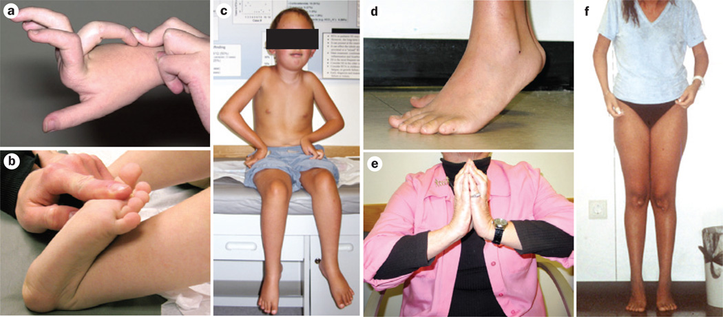
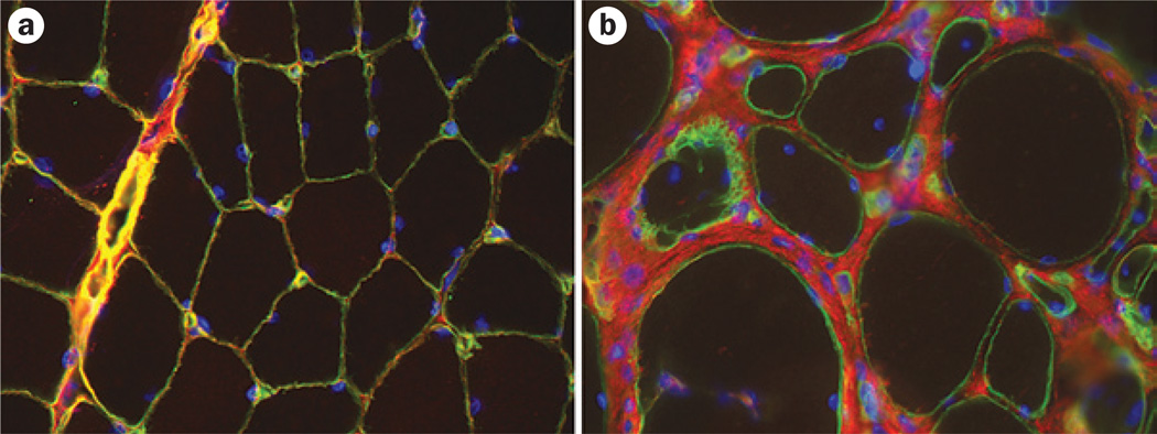
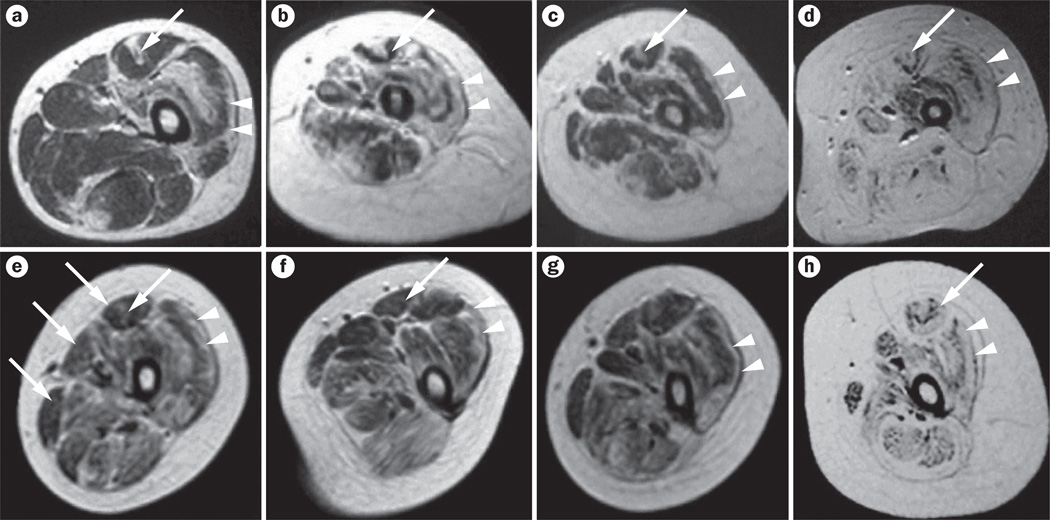
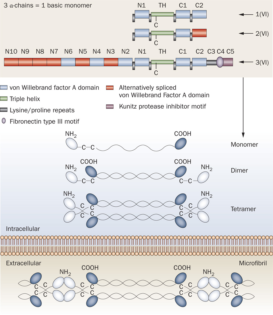
The collagen VI-related myopathy known as Ullrich congenital muscular dystrophy is an early-onset disease that combines substantial muscle weakness with striking joint laxity and progressive contractures. Patients might learn to walk in early childhood; however, this ability is subsequently lost, concomitant with the development of frequent nocturnal respiratory failure. Patients with intermediate phenotypes of collagen VI-related myopathy display a lesser degree of weakness and a longer period of ambulation than do individuals with Ullrich congenital muscular dystrophy, and the spectrum of disease finally encompasses mild Bethlem myopathy, in which ambulation persists into adulthood. Dominant and recessive autosomal mutations in the three major collagen VI genes-COL6A1, COL6A2, and COL6A3-can underlie this entire clinical spectrum, and result in deficient or dysfunctional microfibrillar collagen VI in the extracellular matrix of muscle and other connective tissues, such as skin and tendons. The potential effects on muscle include progressive dystrophic changes, fibrosis and evidence for increased apoptosis, which potentially open avenues for pharmacological intervention. Optimized respiratory management, including noninvasive nocturnal ventilation together with careful orthopedic management, are the current mainstays of treatment and have already led to a considerable improvement in life expectancy for children with Ullrich congenital muscular dystrophy.


Figures

Figure 1
Figure 1
Joint laxity and progressive joint contractures in patients with Ullrich CMD and Bethlem myopathy. a,b | Typical pronounced distal hyperlaxity seen in children with Ullrich CMD, particularly in the fingers. c | A patient with typical Ullrich CMD who had an initial period of ambulation but can no longer walk—note evolving contractures in the shoulders, elbows and knees, round face with mild facial erythema, as well as distal joint laxity. d | Pronounced Achilles tendon contractures in a teenager with Bethlem myopathy. e | Typical long finger flexor contractures in an adult patient with Bethlem myopathy prevent full extension of the fingers when the palms are placed together with dorsiflexed hands and elevated elbows (the ‘Bethlem sign’). f | An adult with Bethlem myopathy. The typical upper body posture results from contractures at the elbows and shoulders. She also has contractures of the Achilles tendons. Abbreviation: CMD, congenital muscular dystrophy. Written consent for publication was obtained from the father of the patient featured in part c.
Figure 2
Figure 2
Immunohistochemical identification of collagen VI in the muscle. Images showing dual immunohistochemical labeling for collagen VI (red) and the basement marker laminin subunit γ-1 (green). a | Note the colocalization of collagen VI and laminin γ-1 in the basement membrane in a healthy individual, which results in a yellow color. b | In a patient with collagen VI-related myopathy, a gap is evident between the collagen VI staining and the basement membrane staining. This patient has a dominant-negative mutation, so that altered collagen VI can be excreted into the matrix but is then not able to function or interact properly.
Figure 3
Figure 3
Muscle MRI findings in patients with collagen VI-related myopathies. T1-weighted MRI scans of the thigh in patients with a–d | Bethlem myopathy and e–h | Ullrich congenital muscular dystrophy of variable severity. Characteristic fatty degeneration (white areas) is seen along the fascia in the center of the rectus femoris (long arrows) and along the rim of the vastus lateralis (arrowheads)—the characteristic ‘outside-in’ pattern. The findings in parts d and h are still visible in the patients with severe forms of these diseases. Permission obtained from Elsevier Ltd © Mercuri, E. Neuromuscul. Disord. 15, 303–310 (2005).
Figure 4
Figure 4
Schematic diagram of the domains and organization of collagen VI. One of each of the three α-chains (α1, α2 and α3) combine along their collagenous triple-helical domains to form the collagen VI monomer. Within the cell, these monomers associate to form dimers, which pair up into tetramers. These tetramers are then secreted into the extracellular matrix, where they assemble by interacting at the globular domains to form the microfibrillar structures typical of collagen VI. Abbreviations: C1–C5, C-terminal globular domains 105; N1–N10, N-terminal globular domains 1–10; TH, triple-helical domain. Modified after Lampe and Bushby (2005) and Furthmayr et al. (1983).

References

    1. Okada M, et al. Primary collagen VI deficiency is the second most common congenital muscular dystrophy in Japan. Neurology. 2007;69:1035–1042. - PubMed
    1. Norwood FL, et al. Prevalence of genetic muscle disease in northern England: in-depth analysis of a muscle clinic population. Brain. 2009;132:3175–3186. - PMC - PubMed
    1. Peat RA, et al. Diagnosis and etiology of congenital muscular dystrophy. Neurology. 2008;71:312–321. - PubMed
    1. Ullrich O. Congenital, atonic–sclerotic muscular dystrophy, an additional type of heredo-degenerative illness of the neuromuscular system [German] Z. Ges. Neurol. Psychiat. 1930;126:171–201.
    1. Ullrich O. Congenital atonic–sclerotic muscular dystrophy [German] Monatsschr. Kinderheilkd. 1930;47:502–510.

Substances