. 2011 Mar;2011(2):31-42.
doi: 10.2147/RRB.S13463.

Duchenne muscular dystrophy gene therapy: Lost in translation?

Affiliations

Duchenne muscular dystrophy gene therapy: Lost in translation?

Dongsheng Duan. Res Rep Biol. 2011 Mar.

Abstract

A milestone of molecular medicine is the identification of dystrophin gene mutation as the cause of Duchenne muscular dystrophy (DMD). Over the last 2 decades, major advances in dystrophin biology and gene delivery technology have created an opportunity to treat DMD with gene therapy. Remarkable success has been achieved in treating dystrophic mice. Several gene therapy strategies, including plasmid transfer, exon skipping, and adeno-associated virus-mediated microdystrophin therapy, have entered clinical trials. However, therapeutic benefit has not been realized in DMD patients. Bridging the gap between mice and humans is no doubt the most pressing issue facing DMD gene therapy now. In contrast to mice, dystrophin-deficient dogs are genetically and phenotypically similar to human patients. Preliminary gene therapy studies in the canine model may offer critical insights that cannot be obtained from murine studies. It is clear that the canine DMD model may represent an important link between mice and humans. Unfortunately, our current knowledge of dystrophic dogs is limited, and the full picture of disease progression remains to be clearly defined. We also lack rigorous outcome measures (such as in situ force measurement) to monitor therapeutic efficacy in dystrophic dogs. Undoubtedly, maintaining a dystrophic dog colony is technically demanding, and the cost of dog studies cannot be underestimated. A carefully coordinated effort from the entire DMD community is needed to make the best use of the precious dog resource. Successful DMD gene therapy may depend on valid translational studies in dystrophin-deficient dogs.


Conflict of interest statement

Disclosure

The author reports no conflicts of interest in this work.

Figures

Figure 1
Figure 1
Schematic illustration of full-length dystrophin, utrophin, and representative minidystrophin and microdystrophin. Abbreviations: N, the N-terminal domain of the dystrophin protein; H1–4, hinges 1–4 in the rod domain of the dystrophin protein; numeric numbers, spectrin-like repeats in the dystrophin rod domain. Positively charged repeats are in white color. Repeats 11–17 represent the second actin-binding domain. CR, the cysteine-rich domain in the dystrophin protein; C, the C-terminal domain of the dystrophin protein; nNOS, neuronal nitric oxide synthase. Empty boxes denote the regions that are deleted in each respective minidystrophin and microdystrophin construct. Among three microgenes listed here, the ΔR3–19/Δ20–21/ΔC microdystrophin has been tested in affected dogs and human patients. However, these trials have failed to demonstrate a therapeutic efficacy. The hinge 2 region in ΔR4–R23/ΔC microdystrophin has been shown to compromise function. The potentially deleterious hinges 2 and 3 are removed in the ΔR2–15/ΔR18–23/ΔC microgene. Further, this microgene carries the nNOS localization domain.
Figure 2
Figure 2
Mouse and dog models of DMD. A) Representative pictures of an 8-month-old dystrophin-deficient mdx4cv mouse (left panel), a 23-month-old mdx mouse (middle two panels), and a 3-month-old myoD/dystrophin double knockout (m-dKO) mouse and a 3-month-old mdx mouse (right panel). Adult dystrophin-null mice do not display clinical symptoms (left panel). The middle two panels show body emaciation and mouse in aged dystrophin-null mouse. (Arrow points to kyphosis seen in aged mdx mice. The left panel is a full-body view. The right-side panel represents a close view of kyphosis after the skin is removed.) The difference in the genetic background results in different fur color in m-dKO (brown) and mdx (black) mice (right panel). Findings from dKO mice may be biased by their mixed genetic background. B) Representative pictures of a 2-year-old normal golden retriever (top) and an affected GRMD (bottom) dog. The affected dog shows body and limb muscle atrophy, drooling, and ulnocarpal joints (forelimbs) dropped to the ground level. C) Representative muscle immunofluorescence staining and histopathology staining in normal (left panels) and GRMD (right panels) dogs. Dystrophin is detected by an antibody (Dys-1) specific for spectrin-like repeats 6–8 (top row). Muscle pathology is revealed by hematoxylin–eosin (HE) staining (middle row) and Masson’s trichrome staining (bottom row). HE staining shows inflammatory cell infiltration, central nucleation, and variable myofiber size. Masson’s trichrome staining shows fibrosis. Blue color in Masson’s trichrome staining images represents fibrotic tissue. Abbreviations: DMD, Duchenne muscular dystrophy; GRMD, golden retriever muscular dystrophy.
Figure 3
Figure 3
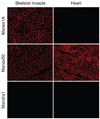
Detection of canine dystrophin with monoclonal antibodies. Monoclonal dystrophin antibodies were used to detect dystrophin expression in a normal dog skeletal muscle and the heart. Representative immunostaining photomicrographs are shown for antibodies Manex1 A, Manex50, and Mandra1. Manex1A recognizes an epitope encoded by exon 1 (N-terminal domain). Interestingly, it only reacts with dystrophin in dog skeletal muscle. Manex50 maps to an epitope encoded by exon 50 (rod domain). This antibody can detect dog dystrophin in both skeletal and cardiac muscles. Mandra1 is supposed to react with an epitope encoded by exon 70 (C-terminal domain). However, it fails to reveal canine dystrophin in either skeletal muscle or the heart.

References

    1. Hoffman EP, Brown RH, Jr, Kunkel LM. Dystrophin: the protein product of the Duchenne muscular dystrophy locus. Cell. 1987;51(6):919–928. - PubMed
    1. Kunkel LM. 2004 William Allan Award address. Cloning of the DMD gene. Am J Hum Genet. 2005;76(2):205–214. - PMC - PubMed
    1. Koenig M, Hoffman EP, Bertelson CJ, Monaco AP, Feener C, Kunkel LM. Complete cloning of the Duchenne muscular dystrophy (DMD) cDNA and preliminary genomic organization of the DMD gene in normal and affected individuals. Cell. 1987;50(3):509–517. - PubMed
    1. Romitti P, Puzhankara S, Mathews K, et al. Centers for Disease Control and Prevention (CDC). Prevalence of Duchenne/Becker muscular dystrophy among males aged 5–24 years – four states, 2007. Morb Mortal Wkly Rep. 2009;58(40):1119–1122. - PubMed
    1. Dooley J, Gordon KE, Dodds L, MacSween J. Duchenne muscular dystrophy: a 30-year population-based incidence study. Clin Pediatr (Phila) 2010;49(2):177–179. - PubMed

LinkOut - more resources