Review
. 2012 Jan;347(1):85-101.
doi: 10.1007/s00441-011-1199-1. Epub 2011 Jun 21.

Deconstructing the mechanisms and consequences of TGF-β-induced EMT during cancer progression

Affiliations
Review

Deconstructing the mechanisms and consequences of TGF-β-induced EMT during cancer progression

Michael K Wendt et al. Cell Tissue Res. 2012 Jan.

Abstract

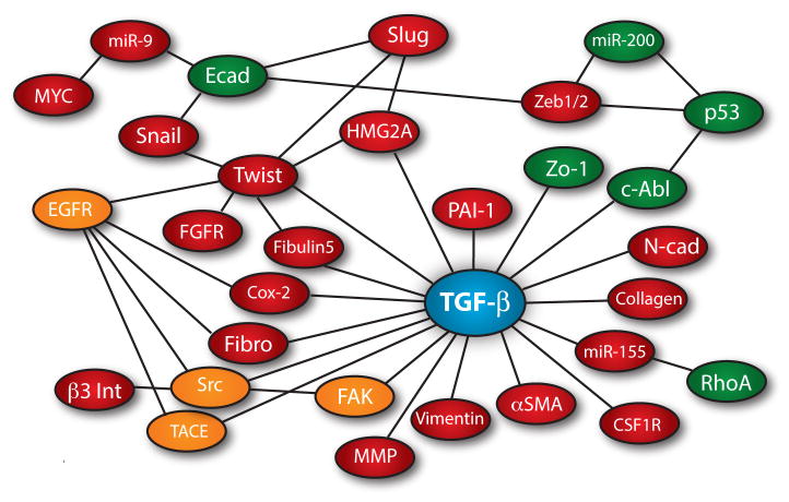
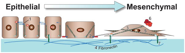
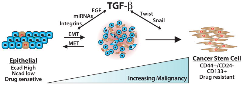
Transforming growth factor-β (TGF-β) is a potent pleiotropic cytokine that regulates mammalian development, differentiation, and homeostasis in essentially all cell types and tissues. TGF-β normally exerts anticancer activities by prohibiting cell proliferation and by creating cell microenvironments that inhibit cell motility, invasion, and metastasis. However, accumulating evidence indicates that the process of tumorigenesis, particularly that associated with metastatic progression, confers TGF-β with oncogenic activities, a functional switch known as the "TGF-β paradox." The molecular determinants governing the TGF-β paradox are complex and represent an intense area of investigation by researchers in academic and industrial settings. Recent findings link genetic and epigenetic events in mediating the acquisition of oncogenic activity by TGF-β, as do aberrant alterations within tumor microenvironments. These events coalesce to enable TGF-β to direct metastatic progression via the stimulation of epithelial-mesenchymal transition (EMT), which permits carcinoma cells to abandon polarized epithelial phenotypes in favor of apolar mesenchymal-like phenotypes. Attempts to deconstruct the EMT process induced by TGF-β have identified numerous signaling molecules, transcription factors, and microRNAs operant in mediating the initiation and resolution of this complex transdifferentiation event. In addition to its ability to enhance carcinoma cell invasion and metastasis, EMT also endows transitioned cells with stem-like properties, including the acquisition of self-renewal and tumor-initiating capabilities coupled to chemoresistance. Here, we review recent findings that delineate the pathophysiological mechanisms whereby EMT stimulated by TGF-β promotes metastatic progression and disease recurrence in human carcinomas.


Figures

Fig. 1
Fig. 1
The EMT signaling network and transcriptome regulated by TGF-β. Red spheres represent genes whose expression is increased, while Green spheres signify genes whose expression is decreased. Orange spheres depict increased enzymatic activity stimulated by TGF-β during EMT. Lines between nodes indicate a described interaction or transactivation between those molecules during EMT.
Fig. 2
Fig. 2
Transdifferentiation from polarized epithelial-like to mesenchymal-like morphologies. Provided numbers depict classical biomarkers of initiated EMT programs, while accompanying boxes describe the EMT-related functions of these highlighted markers. (1) E-cadherin, the primary component of adherens junctions. During EMT, TGF-β represses E-cadherin expression, as well as induces its internalization from the plasma membrane (Miettinen, et al., 1994). (2) N-cadherin, an adhesion molecule that promotes cellular migration (Hazan, et al., 2000). EMT stimulated by TGF-β is associated with increased expression of N-cadherin (Bhowmick, et al., 2001a). (3) Vimentin, an intermediate filament protein that is expressed in all primitive cell types, but not in differentiated epithelial cells. Vimentin may function to drive EMT programs (Mendez, et al., 2010), and also serves as a canonical marker for detecting transdifferentiated mesenchymal cell types (Grunert, et al., 2003). (4) Fibronectin, a critical ECM component whose elevated production by cancer cells is classically associated with EMT programs. TGF-β is a potent inducer of fibronectin production and deposition into the ECM (Ignotz and Massague, 1986). (5) α-Smooth Muscle Actin (α-SMA), a major component of contractile microfilaments and a canonical marker for detecting myofibroblasts. TGF-β stimulation of EMT elicits α-SMA expression (Masszi, et al., 2003), which strongly associates with increased tumor invasion, and with decreased patient survival (Yazhou, et al., 2004). (6) Matrix Metalloproteinases (MMPs), proteolytically degrade basement membranes to allow primary tumor cells to invade the surrounding tissue and intrasvasate into tumor associated vasculature. While several MMPs are induced during EMT, MMP-9 is a well-established target of TGF-β signaling (Duivenvoorden, et al., 1999, Kim, et al., 2007a, Lee, et al., 2008). (7) β3 integrin, a transmembrane protein that physically links the extracellular matrix to intracellular signaling systems and the cytoskeleton via focal adhesion complexes. β3 integrin is rapidly and robustly upregulated by TGF-β and interacts physically with TβR-II via a FAK-dependent mechanism (Galliher and Schiemann, 2006, Wendt and Schiemann, 2009).
Fig. 3
Fig. 3
The consequences of EMT programs induced by TGF-β. Administration of TGF-β readily elicits the formation of a metastable EMT state in normal and malignant cells. The restoration of epithelial phenotypes transpires through mesenchymal-epithelial transitions (METs), which occur normally during developmental EMTs and perhaps during metastatic outgrowth associated with oncogenic EMTs. Prolonged exposure of cells to TGF-β or other EMT-initiating factors supports the continued development and expansion of cancer stem cells, which collectively are chemoresistant and underlie disease recurrence. How, when, and where cancer stems cells undergo MET during secondary tumor outgrowth remains to be determined definitively.

References

    1. Adhikari AS, Agarwal N, Wood BM, Porretta C, Ruiz B, Pochampally RR, Iwakuma T. CD117 and Stro-1 identify osteosarcoma tumor-initiating cells associated with metastasis and drug resistance. Cancer Res. 2010;70:4602–4612. - PMC - PubMed
    1. Al-Hajj M, Wicha MS, Benito-Hernandez A, Morrison SJ, Clarke MF. Prospective identification of tumorigenic breast cancer cells. Proc Natl Acad Sci U S A. 2003;100:3983–3988. - PMC - PubMed
    1. Alexandrow MG, Kawabata M, Aakre M, Moses HL. Overexpression of the c-Myc oncoprotein blocks the growth-inhibitory response but is required for the mitogenic effects of transforming growth factor β1. Proc Natl Acad Sci U S A. 1995;92:3239–3243. - PMC - PubMed
    1. Allington TM, Galliher-Beckley AJ, Schiemann WP. Activated Abl kinase inhibits oncogenic transforming growth factor-β signaling and tumorigenesis in mammary tumors. FASEB J. 2009;23:4231–4243. - PMC - PubMed
    1. Anzano MA, Roberts AB, De Larco JE, Wakefield LM, Assoian RK, Roche NS, Smith JM, Lazarus JE, Sporn MB. Increased secretion of type β transforming growth factor accompanies viral transformation of cells. Mol Cell Biol. 1985;5:242–247. - PMC - PubMed

Publication types

MeSH terms

LinkOut - more resources