. 2011 Nov;31(8):1203-12.
doi: 10.1007/s10571-011-9722-1. Epub 2011 Jun 21.

Isoliquiritigenin is a novel NMDA receptor antagonist in kampo medicine yokukansan

Affiliations

Isoliquiritigenin is a novel NMDA receptor antagonist in kampo medicine yokukansan

Zenji Kawakami et al. Cell Mol Neurobiol. 2011 Nov.

Abstract

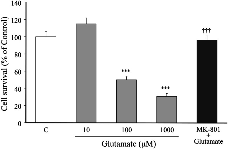
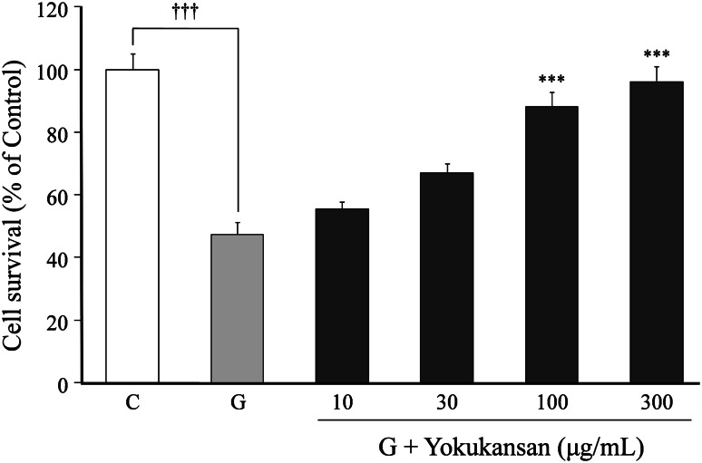
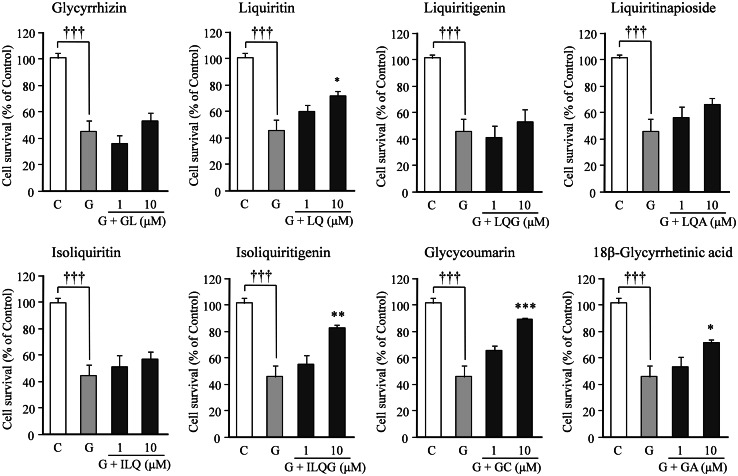
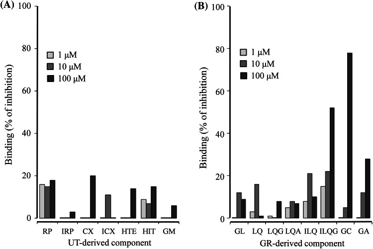
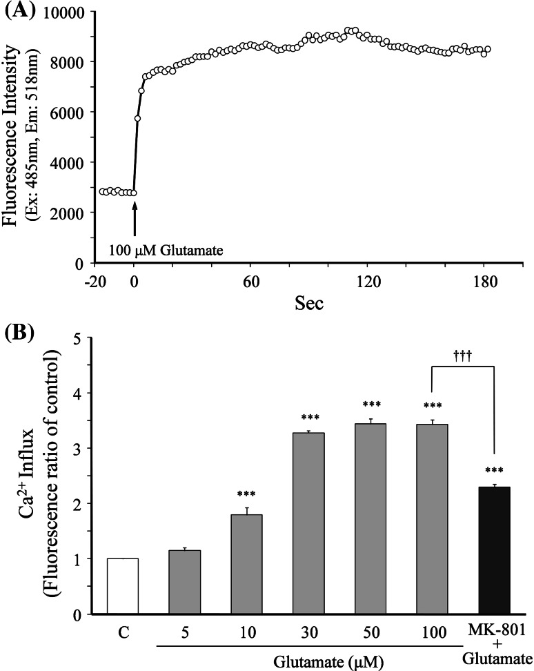
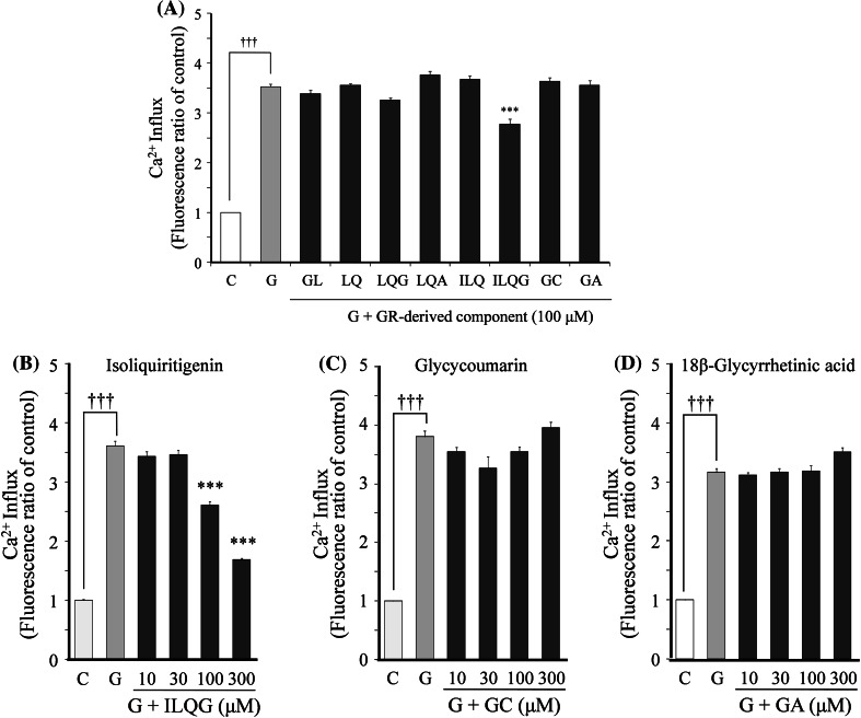
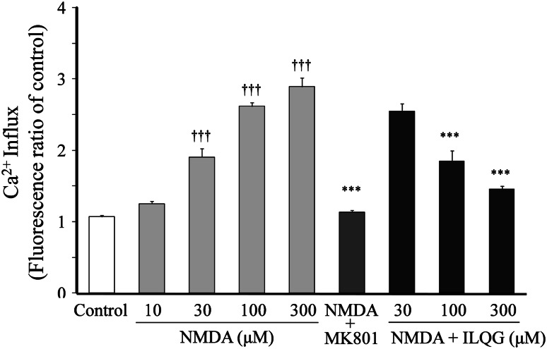
Effects of a traditional Japanese medicine, yokukansan, which is composed of seven medicinal herbs, on glutamate-induced cell death were examined using primary cultured rat cortical neurons. Yokukansan (10-300 μg/ml) inhibited the 100 μM glutamate-induced neuronal death in a concentration-dependent manner. Among seven constituent herbs, higher potency of protection was found in Uncaria thorn (UT) and Glycyrrhiza root (GR). A similar neuroprotective effect was found in four components (geissoschizine methyl ether, hirsuteine, hirsutine, and rhynchophylline) in UT and four components (glycycoumarin, isoliquiritigenin, liquiritin, and 18β-glycyrrhetinic acid) in GR. In the NMDA receptor binding and receptor-linked Ca(2+) influx assays, only isoliquiritigenin bound to NMDA receptors and inhibited the glutamate-induced increase in Ca(2+) influx. Glycycoumarin and 18β-glycyrrhetinic acid bound to NMDA receptors, but did not inhibit the Ca(2+) influx. The four UT-derived components did not bind to NMDA receptors. The present results suggest that neuroprotective components (isoliquiritigenin, glycycoumarin, liquiritin, and 18β-glycyrrhetinic acid in GR and geissoschizine methyl ether, hirsuteine, hirsutine, and rhynchophylline in UT) are contained in yokukansan, and isoliquiritigenin, which is one of them, is a novel NMDA receptor antagonist.


Conflict of interest statement

None.

Figures

Fig. 1
Fig. 1
Relationship between glutamate concentration and neuronal death. Primary cultured neurons were incubated for 24 h in the medium containing various concentrations (10–1,000 μM) of glutamate or 100 μM glutamate + MK-801 (20 μM). The medium for the control did not contain glutamate. Each value calculated as percentage of the MTT activity in control cells is represented as the mean ± SEM (n = 6). ***P < 0.001 versus control (C), and ††† P < 0.001 versus 100 μM glutamate (G): one-way ANOVA + Scheffé’s test
Fig. 2
Fig. 2
Inhibitory effects of yokukansan on 100 μM glutamate-induced cell death. Primary cultured neurons were incubated for 24 h in the medium containing 100 μM glutamate or 100 μM glutamate + various concentrations (10–300 μg/ml) of yokukansan. The control medium did not contain glutamate. Each value calculated as a percentage of the MTT activity in control cells is represented as the mean ± SEM (n = 6). ***P < 0.001 versus control (C) and ††† P < 0.001 versus glutamate (G): one-way ANOVA + Scheffé’s test
Fig. 3
Fig. 3
Effects of seven constituent herbs of yokukansan on glutamate-induced neuronal death. Primary cultured neurons were incubated for 24 h in the medium containing 100 μM glutamate or 100 μM glutamate + one constituent herb (20 μg/ml): Atractylodis lancea rhizome (ALR), Poria sclerotium (PS), Cnidium rhizome (CR), Uncaria thorn (UT), Japanese Angelica root (JAR), Bupleurum root (BR), or Glycyrrhiza root (GR). The control medium did not contain glutamate. Each value calculated as a percentage of the MTT activity in control cells is represented as the mean ± SEM (n = 6). ††† P < 0.001 versus control (C) and **P < 0.01 and ***P < 0.001 versus glutamate (G): one-way ANOVA + Scheffé’s test
Fig. 4
Fig. 4
Effects of seven components of UT on glutamate-induced neuronal death. Primary cultured neurons were incubated for 24 h in medium containing 100 μM glutamate or 100 μM glutamate + one component (1 or 10 μM): rhynchophylline (RP), isorhynchophylline (IRP), corynoxeine (CX), isocorynoxeine (ICX), hirsuteine (HTE), hirsutine (HTI), or geissoschizine methyl ether (GM). The control medium did not contain glutamate. Each value calculated as a percentage of the MTT activity in control cells is represented as the mean ± SEM (n = 6). ††† P < 0.001 versus corresponding control (C), **P < 0.01 and ***P < 0.001 versus glutamate (G): one-way ANOVA + Scheffé’s test
Fig. 5
Fig. 5
Effects of eight components of GR on glutamate-induced neuronal death. Primary cultured neurons were incubated for 24 h in the medium containing 100 μM glutamate or 100 μM glutamate + one component (1 or 10 μM): glycyrrhizin (GL), liquiritin (LQ), liquiritigenin (LQG), liquiritinapioside (LQA), isoliquiritin (ILQ), isoliquitigenin (ILQG), glycycoumarin (GC), or 18β-glycyrretinic acid (GA). The control medium did not contain glutamate. Each value calculated as a percentage of the MTT activity in control cells is represented as the mean ± SEM (n = 6). ††† P < 0.001 versus corresponding control (C), *P < 0.05, **P < 0.01, and ***P < 0.001 versus glutamate (G): one-way ANOVA + Scheffé’s test
Fig. 6
Fig. 6
Competitive binding of UT- (a) and GR-derived components (b) to glutamate recognition sites in NMDA receptors. The membrane fraction of the rat cerebral cortex was used for the binding assay. Binding of each component (1–100 μM) is expressed as percentage inhibition of each component against the total specific binding of a radioligand ([3H]CGP-39653) to glutamate binding site. Each value is expressed as the mean of duplicate determinations. UT-derived components were rhynchophylline (RP), isorhynchophylline (IRP), corynoxeine (CX), isocorynoxeine (ICX), hirsuteine (HTE), hirsutine (HTI), and geissoschizine methyl ether (GM). GR-derived components were glycyrrhizin (GL), liquiritin (LQ), liquiritigenin (LQG), liquiritinapioside (LQA), isoliquiritin (ILQ), isoliquitigenin (ILQG), glycycoumarin (GC), and 18β-glycyrretinic acid (GA)
Fig. 7
Fig. 7
Glutamate-induced increase in Ca2+ influx. Intracellular fluorescent Ca2+ in cultured cortical neurons was measured using Fluo 4 as an indicator. a A typical time–response curve of fluorescence intensity in neurons after 100 μM glutamate was added to the recording buffer. Glutamate was added by an automated dispenser. Fluorescence measurements were taken at 2 s intervals for a total elapsed time of 200 s. b The relationship between the glutamate concentration and Ca2+ influx. The Ca2+ influx induced by glutamate (5–100 μM) was compared at 3 min after addition of glutamate. Each value is represented as the mean ± SEM (n = 6). ***P < 0.001 versus control (C), and ††† P < 0.001 versus 100 μM glutamate: one-way ANOVA + Scheffé’s test
Fig. 8
Fig. 8
Effects of GR-derived components on glutamate-induced increase in Ca2+ influx. a The effects of eight components on 100 μM glutamate-induced Ca2+ influx. bd The effects of concentration of isoliquiritigenin, glycycoumarin, and 18β-glycyrrhetinic acid on the glutamate-induced increase in Ca2+ influx. The effect of each component on the Ca2+ influx was evaluated at 3 min after addition of glutamate. Each value expressed as the fluorescence ratio of the control is represented as the mean ± SEM (n = 6). ††† P < 0.001 versus control (C) and ***P < 0.001 versus glutamate (G): one-way ANOVA + Scheffé’s test. Glycyrrhiza root-derived components were glycyrrhizin (GL), liquiritin (LQ), liquiritigenin (LQG), liquiritinapioside (LQA), isoliquiritin (ILQ), isoliquitigenin (ILQG), glycycoumarin (GC), and 18β-glycyrretinic acid (GA)
Fig. 9
Fig. 9
Effects of isoliquilitigenin (ILQG) on NMDA-induced increase in Ca2+ influx. The Ca2+ influx was measured using Fluo 4 as an indicator. The Ca2+ influx induced by NMDA (10–300 μM) was compared at 3 min after addition of NMDA. The effects of MK-801 (1 μM) and ILQG (30–300 μM) on the Ca2+ influx induced by NMDA were evaluated at 3 min after addition of 300 μM NMDA. Each value expressed as the fluorescence ratio of the control is represented as the mean ± SEM (n = 6). ††† P < 0.001 versus control and ***P < 0.001 versus NMDA (300 μM): one-way ANOVA + Scheffé’s test

References

    1. Arimatsu Y, Hatanaka H (1986) Estrogen treatment enhances survival of cultured fetal rat amygdala neurons in a defined medium. Brain Res 391:151–159 - PubMed
    1. Bruno V, Copani A, Knöpfel T, Kuhn R, Casabona G, Dell’Albani P, Condorelli DF, Nicoletti F (1995) Activation of metabotropic glutamate receptors coupled to inositol phospholipid hydrolysis amplifies NMDA-induced neuronal degeneration in cultured cortical cells. Neuropharmacology 34:1089–1098 - DOI - PubMed
    1. Cho SC, Rhim JH, Son YH, Lee SJ, Park SC (2010) Suppression of ROS generation by 4, 4-diaminodiphenylsulfone in non-phagocytic human diploid fibroblasts. Exp Mol Med 42:223–232 - DOI - PMC - PubMed
    1. Edwards MA, Loxley RA, Williams AJ, Connor M, Phillips JK (2007) Lack of functional expression of NMDA receptors in PC12 cells. Neurotoxicology 28:876–885 - DOI - PubMed
    1. Froissard P, Duval D (1994) Cytotoxic effects of glutamic acid on PC12 cells. Neurochem Int 24:485–493 - DOI - PubMed

MeSH terms

LinkOut - more resources