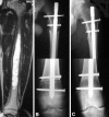
Intercalary femur allografts are an acceptable alternative after tumor resection
- PMID: 21691906
- PMCID: PMC3270162
- DOI: 10.1007/s11999-011-1952-5
Intercalary femur allografts are an acceptable alternative after tumor resection
Abstract
Background: With the improved survival for patients with malignant bone tumors, there is a trend to reconstruct defects using biologic techniques. While the use of an intercalary allograft is an option, the procedures are technically demanding and it is unclear whether the complication rates and survival are similar to other approaches.
Questions/purposes: We evaluated survivorship, complications, and functional scores of patients after receiving intercalary femur segmental allografts.
Patients and methods: We retrospectively reviewed 83 patients who underwent an intercalary femur segmental allograft reconstruction. We determined allograft survival using the Kaplan-Meier method. We evaluated patient function with the Musculoskeletal Tumor Society scoring system. Minimum followup was 24 months (median, 61 months; range, 24-182 months).
Results: Survivorship was 85% (95% confidence interval: 93%-77%) at 5 years and 76% (95% confidence interval: 89%-63%) at 10 years. Allografts were removed in 15 of the 83 patients: one with infection, one with local recurrence, and 13 with fractures. Of the 166 host-donor junctions, 22 (13%) did not initially heal. Nonunion rate was 19% for diaphyseal junctions and 3% for metaphyseal junctions. We observed an increase in the diaphysis nonunion rate in patients fixed with nails (28%) compared to those fixed with plates (15%). Fracture rate was 17% and related to areas of the allograft not adequately protected with internal fixation. All patients without complications had mainly good or excellent Musculoskeletal Tumor Society functional results.
Conclusions: Diaphyseal junctions have higher nonunion rates than metaphyseal junctions. The internal fixation should span the entire allograft to avoid the risk of fracture. Our observations suggest segmental allograft of the femur provides an acceptable alternative in reconstructing tumor resections.
Level of evidence: Level IV, therapeutic study. See the Guidelines for Authors for a complete description of levels of evidence.
Figures


Similar articles
-
Does the Addition of a Vascularized Fibula Improve the Results of a Massive Bone Allograft Alone for Intercalary Femur Reconstruction of Malignant Bone Tumors in Children?Clin Orthop Relat Res. 2021 Jun 1;479(6):1296-1308. doi: 10.1097/CORR.0000000000001639. Clin Orthop Relat Res. 2021. PMID: 33497066 Free PMC article.
-
Do Massive Allograft Reconstructions for Tumors of the Femur and Tibia Survive 10 or More Years after Implantation?Clin Orthop Relat Res. 2020 Mar;478(3):517-524. doi: 10.1097/CORR.0000000000000806. Clin Orthop Relat Res. 2020. PMID: 32168064 Free PMC article.
-
What Is the Survival of the Telescope Allograft Technique to Augment a Short Proximal Femur Segment in Children After Resection and Distal Femur Endoprosthesis Reconstruction for a Bone Sarcoma?Clin Orthop Relat Res. 2021 Aug 1;479(8):1780-1790. doi: 10.1097/CORR.0000000000001686. Clin Orthop Relat Res. 2021. PMID: 33635286 Free PMC article.
-
Hemicortical resection and inlay allograft reconstruction for primary bone tumors: a retrospective evaluation in the Netherlands and review of the literature.J Bone Joint Surg Am. 2015 May 6;97(9):738-50. doi: 10.2106/JBJS.N.00948. J Bone Joint Surg Am. 2015. PMID: 25948521 Review.
-
Intercalary frozen autografts for reconstruction of bone defects following meta-/diaphyseal tumor resection at the extremities.BMC Musculoskelet Disord. 2022 Sep 30;23(1):890. doi: 10.1186/s12891-022-05840-6. BMC Musculoskelet Disord. 2022. PMID: 36180843 Free PMC article. Review.
Cited by
-
An algorithm for surgical treatment of children with bone sarcomas of the extremities.SICOT J. 2024;10:38. doi: 10.1051/sicotj/2024033. Epub 2024 Oct 4. SICOT J. 2024. PMID: 39364963 Free PMC article.
-
Does liquid nitrogen recycled autograft for treatment of bone sarcoma impact local recurrence rate? A systematic review.J Bone Oncol. 2024 Aug 8;48:100628. doi: 10.1016/j.jbo.2024.100628. eCollection 2024 Oct. J Bone Oncol. 2024. PMID: 39257651 Free PMC article.
-
Structural allograft reconstruction of the foot and ankle after tumor resections.Musculoskelet Surg. 2016 Aug;100(2):149-56. doi: 10.1007/s12306-016-0413-4. Epub 2016 Jun 20. Musculoskelet Surg. 2016. PMID: 27324025
-
Outcomes of Intercalary Endoprostheses as a Treatment for Metastases in the Femoral and Humeral Diaphysis.Curr Oncol. 2022 May 13;29(5):3519-3530. doi: 10.3390/curroncol29050284. Curr Oncol. 2022. PMID: 35621674 Free PMC article.
-
Treatment of critical-sized bone defects: clinical and tissue engineering perspectives.Eur J Orthop Surg Traumatol. 2018 Apr;28(3):351-362. doi: 10.1007/s00590-017-2063-0. Epub 2017 Oct 28. Eur J Orthop Surg Traumatol. 2018. PMID: 29080923 Review.
References
-
- Abudu A, Carter SR, Grimer RJ. The outcome and functional results of diaphyseal endoprostheses after tumour excision. J Bone Joint Surg Br. 1996;78:652–657. - PubMed
-
- Canadell J, Forriol F, Cara JA. Removal of metaphyseal bone tumours with preservation of the epiphysis: physeal distraction before excision. J Bone Joint Surg Br. 1994;76:127–132. - PubMed
MeSH terms
LinkOut - more resources
Full Text Sources
Medical
Research Materials

