. 2011 Aug 3;133(30):11795-801.
doi: 10.1021/ja2045925. Epub 2011 Jul 8.

Hyperpolarized [1-13C]-ascorbic and dehydroascorbic acid: vitamin C as a probe for imaging redox status in vivo

Affiliations
Free PMC article

Hyperpolarized [1-13C]-ascorbic and dehydroascorbic acid: vitamin C as a probe for imaging redox status in vivo

Sarah E Bohndiek et al. J Am Chem Soc. .
Free PMC article

Abstract

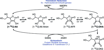
Dynamic nuclear polarization (DNP) of (13)C-labeled metabolic substrates in vitro and their subsequent intravenous administration allow both the location of the hyperpolarized substrate and the dynamics of its subsequent conversion into other metabolic products to be detected in vivo. We report here the hyperpolarization of [1-(13)C]-ascorbic acid (AA) and [1-(13)C]-dehydroascorbic acid (DHA), the reduced and oxidized forms of vitamin C, respectively, and evaluate their performance as probes of tumor redox state. Solution-state polarization of 10.5 ± 1.3% was achieved for both forms at pH 3.2, whereas at pH 7.0, [1-(13)C]-AA retained polarization of 5.1 ± 0.6% and [1-(13)C]-DHA retained 8.2 ± 1.1%. The spin-lattice relaxation times (T(1)'s) for these labeled nuclei are long at 9.4 T: 15.9 ± 0.7 s for AA and 20.5 ± 0.9 s for DHA. Extracellular oxidation of [1-(13)C]-AA and intracellular reduction of [1-(13)C]-DHA were observed in suspensions of murine lymphoma cells. The spontaneous reaction of DHA with the cellular antioxidant glutathione was monitored in vitro and was approximately 100-fold lower than the rate observed in cell suspensions, indicating enzymatic involvement in the intracellular reduction. [1-(13)C]-DHA reduction was also detected in lymphoma tumors in vivo. In contrast, no detectable oxidation of [1-(13)C]-AA was measured in the same tumors, consistent with the notion that tumors maintain a reduced microenvironment. This study demonstrates that hyperpolarized (13)C-labeled vitamin C could be used as a noninvasive biomarker of redox status in vivo, which has the potential to translate to the clinic.


Figures

Figure 1
Figure 1
Primary intracellular oxidation, reduction, and degradation pathways for vitamin C. Enzymes involved in GSH- and NADPH-dependent reduction of dehydroascorbic acid (DHA) are highlighted, with the primary cytosolic enzymes in bold.(14) Abbreviations: AA, ascorbic acid; AFR, ascorbate free radical (also known as semidehydroascorbic acid); DHA, dehydroascorbic acid; DKG, diketogulonic acid; GSH, reduced glutathione; GSSG, oxidized glutathione.
Figure 2
Figure 2
Mechanism of vitamin C transport, in both reduced (AA) and oxidized (DHA) forms, by sodium-dependent vitamin C transporters 1 and 2 (SVCT 1, 2) and glucose transporters (primarily GLUT1, 3 and also GLUT 4), respectively. This is dominated in cancer cells by GLUT1 transport of DHA, with SVCTs either not expressed or having low activity due to the acidic extracellular pH in tumors.,
Figure 3
Figure 3
(a) 13C NMR spectra of hyperpolarized [1-13C]-DHA (175 ppm) at the peak of [1-13C]-AA (179 ppm) production following reaction of 5 mM [1-13C]-DHA with 1, 5, 10, 25, and 50 mM glutathione. Peaks at 173.5 and 175.4 ppm are from diketogulonic acid and an unknown product, respectively. (b) Time course of [1-13C]-AA peak intensity at various glutathione concentrations, normalized to the maximum [1-13C]-DHA intensity. (c) Correlation of the rate of [1-13C]-AA production, [DHA] × kDHA, with the rate of AA production measured spectrophotometrically at equivalent concentrations of DHA and glutathione.
Figure 4
Figure 4
Oxidation of [1-13C]-AA (a, b, e) and reduction of [1-13C]-DHA (c, d, f) in EL4 lymphoma cell suspensions. Hyperpolarized substrates were added to either RPMI medium alone (a, c) or a suspension of 108 EL4 lymphoma cells in RPMI medium. (b) Spectrum obtained at 2.5 min post cell resuspension and (d) at 1.5 min post cell resuspension. Only every fourth spectrum is displayed in the interests of clarity (e, f; time course over 46 s). Peaks at 172.6 and 176.4 ppm are from diketogulonic acid and the C3 carbon of AA,(50) respectively.
Figure 5
Figure 5
13C MR spectra acquired from EL4 murine lymphoma tumors following injection of 30 mM [1-13C]-AA (a) and [1-13C]-DHA (b; every second spectrum shown for clarity). Sequential spectra were collected from a 6-mm tumor slice starting at 15 s post injection over a period of 16 s (a) and 32 s (b). Whereas there was no observable flux of hyperpolarized 13C label from [1-13C]-AA (179 ppm) to [1-13C]-DHA (175 ppm) (a), significant flux was observed from [1-13C]-DHA to [1-13C]-AA, indicating rapid reduction of the injected [1-13C]-DHA in the tumor.

References

    1. Kurhanewicz J.; Vigneron D. B.; Brindle K.; Chekmenev E. Y.; Comment A.; Cunningham C. H.; Deberardinis R. J.; Green G. G.; Leach M. O.; Rajan S. S.; Rizi R. R.; Ross B. D.; Warren W. S.; Malloy C. R. Neoplasia 2011, 13, 81–97. - PMC - PubMed
    1. Bohndiek S. E.; Kettunen M. I.; Hu D. E.; Witney T. H.; Kennedy B. W. C.; Gallagher F. A.; Brindle K. M. Mol. Cancer Ther. 2010, 9, 3278–88. - PMC - PubMed
    1. Day S. E.; Kettunen M. I.; Gallagher F. A.; Hu D. E.; Lerche M.; Wolber J.; Golman K.; Ardenkjaer-Larsen J. H.; Brindle K. M. Nat. Med. 2007, 13, 1382–7. - PubMed
    1. Albers M. J.; Bok R.; Chen A. P.; Cunningham C. H.; Zierhut M. L.; Zhang V. Y.; Kohler S. J.; Tropp J.; Hurd R. E.; Yen Y. F.; Nelson S. J.; Vigneron D. B.; Kurhanewicz J. Cancer Res. 2008, 68, 8607–15. - PMC - PubMed
    1. Keshari K. R.; Wilson D. M.; Chen A. P.; Bok R.; Larson P. E.; Hu S.; Van Criekinge M.; Macdonald J. M.; Vigneron D. B.; Kurhanewicz J. J. Am. Chem. Soc. 2009, 131, 17591–6. - PMC - PubMed

Publication types