Review
. 2011 Oct;102(10):1749-55.
doi: 10.1111/j.1349-7006.2011.02011.x. Epub 2011 Jul 21.

Interface between normal and transformed epithelial cells: a road to a novel type of cancer prevention and treatment

Affiliations
Review

Interface between normal and transformed epithelial cells: a road to a novel type of cancer prevention and treatment

Yasuyuki Fujita. Cancer Sci. 2011 Oct.

Abstract

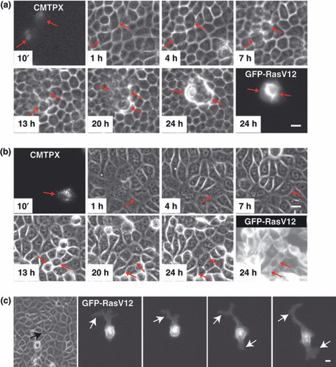
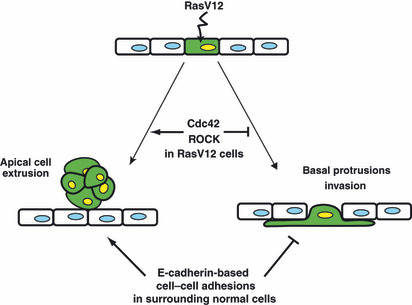
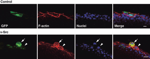
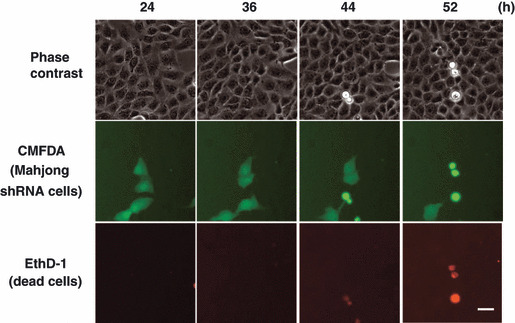
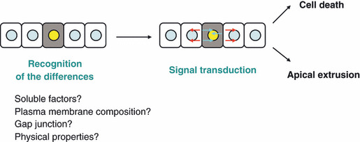
Cell transformation arises from activation of oncoproteins and ⁄ or inactivation of tumor suppressor proteins. During the initial stage of carcinogenesis, transformation occurs in a single cell within an epithelial monolayer. However, it is not known what happens at the interface between normal and transformed cells once the initial transformation has occurred. Using elaborate cell culture systems, recent reports have shown that interactions between normal and transformed epithelial cells can induce various phenomena. For example, when Ras- or Src-transformed cells are surrounded by normal epithelial cells, multiple signaling pathways are activated and the transformed cells are apically extruded from the epithelium. In addition, normal and certain types of transformed cells compete with each other for cell survival, and the transformed cells undergo apoptosis. Importantly, when transformed cells alone are present, neither apoptosis nor elimination from epithelia occurs, indicating that the presence of surrounding normal cells influences the signaling pathways and fate of transformed cells. Comparable phenomena are also observed in zebrafish and mice in vivo model systems. In this review, I will introduce this newly emerging research field and discuss how these studies can potentially lead to establishment of novel types of cancer prevention and treatment.


Figures

Figure 1
Figure 1
Apical extrusion and basal protrusion formation of RasV12‐expressing cells surrounded by normal epithelial cells. Fluorescently labeled MDCK‐pTR GFP‐RasV12 cells (CMTPX) are mixed with non‐transformed MDCK cells (a,c) or MDCK‐pTR GFP‐RasV12 cells (b). Images are extracted from a representative time‐lapse analysis. Scale bar = 20 μm. (a,b) Red arrows indicate fluorescently labeled RasV12 cells. (c) Images are captured at 50‐min intervals. Black arrow and white arrows indicate RasV12 cell surrounded by non‐transformed cells and protrusions, respectively. (Adapted from Hogan et al., with permission, 1, 3.)
Figure 2
Figure 2
Schematic model for the fate of RasV12 cells surrounded by normal epithelial cells. When expression of RasV12 is induced in a single epithelial cell (green) within a monolayer of normal cells, two independent phenomena can occur in a non‐cell‐autonomous fashion. RasV12‐expressing cells are either apically extruded from the monolayer or form basal protrusions beneath the surrounding neighbors. The fate of RasV12 cells is influenced by the activity of Cdc42 and ROCK in RasV12 cells and by E‐cadherin‐based cell–cell adhesions in the surrounding normal cells. (Adapted from Hogan et al., with permission, fig. 5.)
Figure 3
Figure 3
Apical extrusion of a v‐Src‐expressing cell from a monolayer of the enveloping layer in a zebrafish embryo. Immunofluorescence images (semilateral view) of zebrafish embryos (at 8–9 h postfertilization), injected with the GFP‐ (control) or v‐Src‐expressing vector at the 1‐ to 2‐cell stage. Embryos are stained with phalloidin (red) and Hoechst (blue). Arrowhead and arrow indicate the v‐Src‐expressing cell with increased cell height and extruded v‐Src‐expressing cell, respectively. Scale bar = 10 μm. (Adapted from Kajita et al., with permission, fig. 3.)
Figure 4
Figure 4
Cell competition induced by knockdown of Mahjong in MDCK epithelial cells. MDCK‐pTR Mahjong shRNA cells are fluorescently labeled with green fluorescent dye CMFDA (green) and mixed with normal MDCK cells at a ratio of 1:10, and cultured in the presence of tetracycline and EthD‐1 (red) for the indicated times. Scale bar = 30 μm. (Adapted from Tamori et al., with permission, fig. 3.)
Figure 5
Figure 5
Schematic model for the interaction between normal and transformed epithelial cells.

References

    1. Foty RA, Steinberg MS. The differential adhesion hypothesis: a direct evaluation. Dev Biol 2005; 278: 255–63. - PubMed
    1. Krieg M, Arboleda‐Estudillo Y, Puech PH et al. Tensile forces govern germ‐layer organization in zebrafish. Nat Cell Biol 2008; 10: 429–36. - PubMed
    1. Steinberg MS, Takeichi M. Experimental specification of cell sorting, tissue spreading, and specific spatial patterning by quantitative differences in cadherin expression. Proc Natl Acad Sci USA 1994; 91: 206–9. - PMC - PubMed
    1. Hogan C, Dupre‐Crochet S, Norman M et al. Characterization of the interface between normal and transformed epithelial cells. Nat Cell Biol 2009; 11: 460–7. - PubMed
    1. Kajita M, Hogan C, Harris AR et al. Interaction with surrounding normal epithelial cells influences signalling pathways and behaviour of Src‐transformed cells. J Cell Sci 2010; 123: 171–80. - PMC - PubMed

Publication types

Substances

LinkOut - more resources