. 2011 Jul 15;124(Pt 14):2501-10.
doi: 10.1242/jcs.084558. Epub 2011 Jun 21.

TGFB1 disrupts the angiogenic potential of microvascular endothelial cells of the corpus luteum

Affiliations

TGFB1 disrupts the angiogenic potential of microvascular endothelial cells of the corpus luteum

Dulce Maroni et al. J Cell Sci. .

Abstract

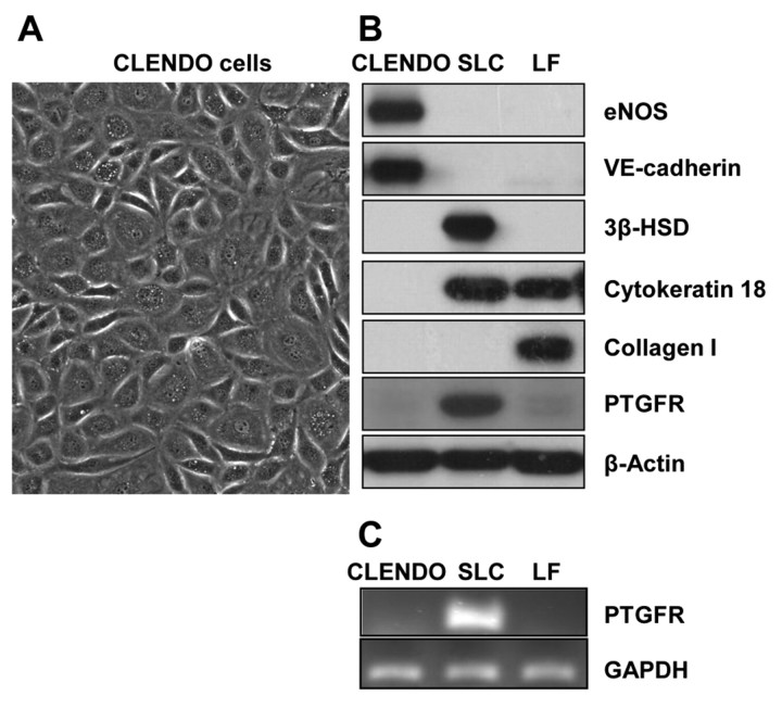
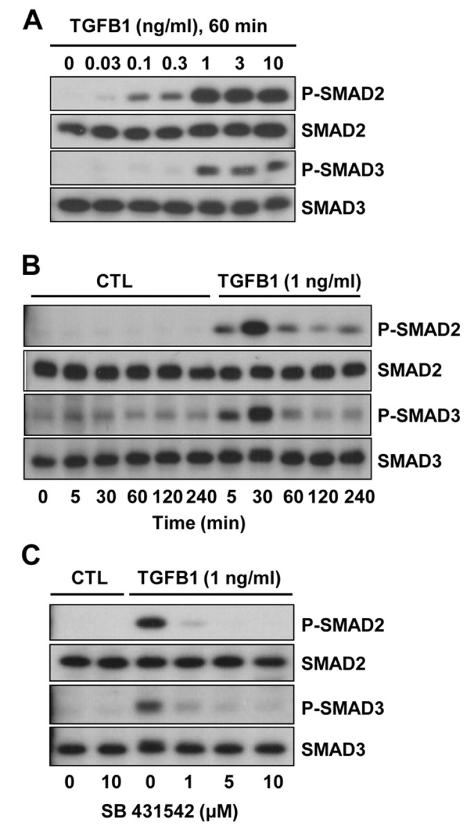
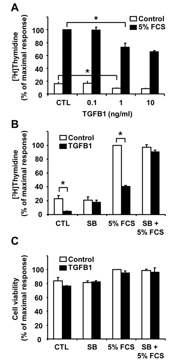
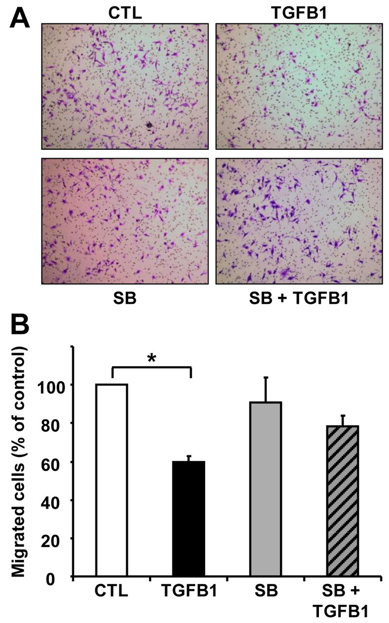
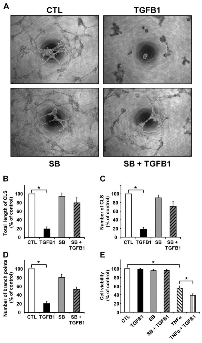
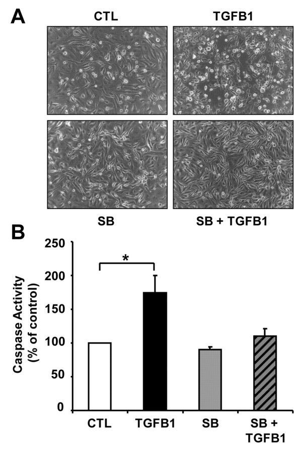
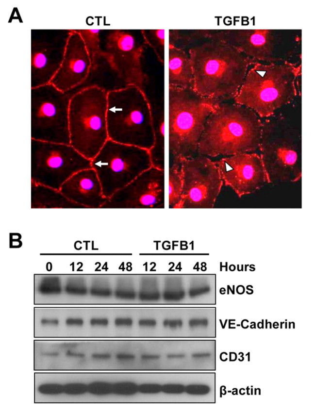
Cyclical formation and regression of the ovarian corpus luteum is required for reproduction. During luteal regression, the microvasculature of the corpus luteum is extensively disrupted. Prostaglandin F2α, a primary signal for luteal regression, induces the expression of transforming growth factor β1 (TGFB1) in the corpus luteum. This study determined the actions of TGFB1 on microvascular endothelial cells isolated from the bovine corpus luteum (CLENDO cells). We hypothesized that TGFB1 participates in the disruption of the microvasculature during luteal regression. TGFB1 activated the canonical SMAD signaling pathway in CLENDO cells. TGFB1 (1 ng/ml) significantly reduced both basal and fetal-calf-serum-stimulated DNA synthesis, without reducing cell viability. TGFB1 also significantly reduced CLENDO cell transwell migration and disrupted the formation of capillary-like structures when CLENDO cells were plated on Matrigel. By contrast, CLENDO cells plated on fibrillar collagen I gels did not form capillary-like structures and TGFB1 induced cell death. Additionally, TGFB1 caused loss of VE-cadherin from cellular junctions and loss of cell-cell contacts, and increased the permeability of confluent CLENDO cell monolayers. These studies demonstrate that TGFB1 acts directly on CLENDO cells to limit endothelial cell function and suggest that TGFB1 might act in the disassembly of capillaries observed during luteal regression.


Figures

Fig. 1.
Fig. 1.
Characterization of CLENDO cells. Microvascular endothelial cells were isolated from bovine corpus luteum and characterized by their morphology and expression of cell markers. (A) CLENDO cells displayed cobblestone morphology. CLENDO cells were characterized by western blot of cell lysates (B) and PCR analysis (C). (B) CLENDO expressed the endothelial cell markers VE-cadherin and eNOS, and did not express cytokeratin 18, PTGFR and the specific markers of steroidogenic luteal cells (SLCs) – HSD3B (3β-HSD) – and of luteal fibroblasts (LFs) – collagen I. (C) The receptor for PGF2α, PTGFR, was detected in SLCs, but was not detectable in CLENDO cells or LFs by PCR analysis.
Fig. 2.
Fig. 2.
TGFB1 induces phosphorylation of SMAD2 and SMAD3 in CLENDO cells. The time-course response (A) and concentration response (B) to TGFB1 are shown. (A) Cells were serum starved and treated with TGFB1 (0–10 ng/ml) for 60 minutes under serum-free conditions. (B) Cells were serum starved and treated with TGFB1 (1 ng/ml) for up to 4 hours under serum-free conditions. (C) Some cells were pretreated (30 minutes) with the selective TGFBR1 kinase inhibitor SB-431542 (0–10 μM) prior to addition of TGFB1 (1 ng/ml) for 60 minutes. Levels of phosphorylated SMAD2 and SMAD3 (P-SMAD2 and P-SMAD3) and total SMAD2 and SMAD3 were determined by western blot analysis. CTL, control.
Fig. 3.
Fig. 3.
TGFB1 reduces DNA synthesis in CLENDO cells. (A) [3H]-thymidine incorporation assay of cells plated at low density and treated with or without TGFB1 (0–10 ng/ml) in the absence (CTL, control) or presence of 5% FCS for 24 hours. (B) [3H]-thymidine incorporation assay of cells plated at low density and treated for 24 hours with or without TGFB1 (1 ng/ml) in the absence (CTL) or presence of 5% FCS. Some cells were pretreated (30 minutes) with the selective TGFBR1 receptor kinase inhibitor SB-431542 (SB, 1 μM) prior to addition of TGFB1. Data are expressed as the percent incorporation of [3H]-thymidine compared to the maximal response group. Data shown represent three independent experiments each performed in triplicate (mean + s.e.m., n=3, *P<0.05). (C) MTT assay of CLENDO cells plated on plastic at low density and treated for 24 hours with or without TGFB1 (1 ng/ml) in the absence (CTL) or presence of 5% FCS. Some cells were pretreated (30 minutes) with SB-431542 (1 μM) prior to addition of TGFB1. Results are expressed as the percent absorbance observed in the control group. Data shown represent three independent experiments each performed in triplicate (mean ± s.e.m., n=3).
Fig. 4.
Fig. 4.
TGFB1 reduces migration of CLENDO cells. CLENDO cells were pretreated with or without SB-431542 (SB, 1 μM) for 30 minutes followed by treatment with TGFB1 (1 ng/ml). Next, cells were plated on transwells and assessed for migration after 6 hours, as described in the Materials and Methods. (A) CLENDO cells found on the bottom of the transwell membrane were fixed, stained with crystal violet and images were obtained for quantification. (B) Quantification of cell migration. Cell migration is represented as a percentage of the number of cells migrated in the control treatment group. Data shown represent five independent experiments each performed in triplicate (mean + s.e.m., n=5, *P<0.05).
Fig. 5.
Fig. 5.
TGFB1 disrupts formation of CLENDO capillary-like structures (CLSs). (A) To perform capillary morphogenesis assays, CLENDO cells were plated (5×10⁴ per well) in growth medium on 48-well plates coated with Matrigel (0.15 ml; 8 mg/ml). Cells were pretreated with or without SB-431542 (SB, 1 μM) for 30 minutes followed by treatment with TGFB1 (1 ng/ml). Pictures were taken under a phase-contrast microscope after 8 hours of incubation at 37°C. CLS formation was quantified as (B) the total length of CLS, (C) the number of CLS and (D) the number of CLS branch points per low-power field. Results are presented as a percentage of measurements obtained in the controls (CTL). Data shown represent four independent experiments each performed in duplicate with similar results (mean ± s.e.m., n=4, *P<0.001). (E) MTT assay of CLENDO cells plated on Matrigel and treated for 24 hours with or without TGFB1 (1 ng/ml) or TNFα (50 ng/ml). Some cells were pretreated (30 minutes) with SB-431542 (1 μM) prior to addition of TGFB1. Data are expressed as the percent cell viability compared to the control group. Data shown represent three independent experiments each performed in triplicate (mean + s.e.m., n=3, *P<0.05).
Fig. 6.
Fig. 6.
Effect of TGFB1 on CLENDO viability when plated on fibrillar collagen type I. (A) Morphology of CLENDO cells after plating (5×10⁴ per well) in growth medium on 48-well plates coated with fibrillar collagen type I gels (0.15 ml; ~2.4 mg/ml). Plated cells were pretreated with or without SB-431542 (SB, 1 μM) for 30 minutes followed by control media (CTL) or TGFB1 (1 ng/ml). Pictures were taken under a phase-contrast microscope after 48 hours incubation at 37°C. (B) Caspase-3 and caspase-7 activity was measured after 24 hours of treatment using the Caspase–Glo 3/7 assay kit. Data are expressed as a percentage of the caspase activity observed in controls. Data shown represent three independent experiments each performed in triplicate with similar results (mean + s.e.m., n=3, *P<0.05).
Fig. 7.
Fig. 7.
TGFB1 causes loss of VE-cadherin from cellular junctions. (A) Confluent monolayers of CLENDO cells were pretreated with or without SB-431542 (1 μM) for 30 minutes followed by treatment with control medium (CTL) or TGFB1 (1 ng/ml) for 48 hours. VE-cadherin expression (red) was determined by immunofluorescence. In control-treated cells, VE-cadherin was localized at cell–cell contacts (arrows). In TGFB1-treated cells, VE-cadherin localization at cellular junctions was irregular (arrowheads) and gaps between cells appeared in the confluent monolayer (arrowheads). (B) The expression of the endothelial cell markers VE-cadherin, eNOS and CD31 was examined by western blotting. β-actin was used as a loading control.
Fig. 8.
Fig. 8.
TGFB1 increases the permeability of CLENDO cell monolayers. CLENDO cells were grown to confluence on membrane inserts (3 μm pore). Cells were pretreated with or without SB-431542 (SB, 1 μM) and then treated with control medium (CTL) or TGFB1 (1 ng/ml) for 24 hours. (A) Monolayer permeability was measured by determining the fluorescence of the bottom chamber medium at the indicated times after the addition of the fluorescent tracer molecule FITC–dextran to the top chamber. Data represent mean ± s.e.m. of fluorescence from a representative experiment performed in triplicate. (B) At the end of the assay, monolayers were fixed and stained with crystal violet to show equivalent density of CLENDO monolayers.

Similar articles

Cited by

References

    1. Alghisi G. C., Ponsonnet L., Rüegg C. (2009). The integrin antagonist cilengitide activates alphaVbeta3, disrupts VE-cadherin localization at cell junctions and enhances permeability in endothelial cells. PLoS ONE 4, e4449. - PMC - PubMed
    1. Alvarez D. F., Huang L., King J. A., ElZarrad M. K., Yoder M. C., Stevens T. (2008). Lung microvascular endothelium is enriched with progenitor cells that exhibit vasculogenic capacity. Am J. Physiol. Lung Cell. Mol. Physiol. 294, L419-L430. - PubMed
    1. Behzadian M. A., Wang X. L., Windsor L. J., Ghaly N., Caldwell R. B. (2001). TGF-β increases retinal endothelial cell permeability by increasing MMP-9: possible role of glial cells in endothelial barrier function. Invest. Ophthalmol. Vis. Sci. 42, 85385-85389. - PubMed
    1. Bein K., Odell-Fiddler E. T., Drinane M. (2004). Role of TGF-beta1 and JNK signaling in capillary tube patterning. Am. J. Physiol. Cell Physiol. 287, C1012-C1022. - PubMed
    1. Berisha B., Meyer H. H. D., Schams D. (2010). Effect of prostaglandin F2 Alpha on local luteotropic and angiogenic factors during induced functional luteolysis in the bovine corpus luteum. Biol. Reprod. 82, 940-947. - PubMed

Publication types

Substances