. 2011 Jul 12;108(28):11452-7.
doi: 10.1073/pnas.1106083108. Epub 2011 Jun 21.

R-spondins function as ligands of the orphan receptors LGR4 and LGR5 to regulate Wnt/beta-catenin signaling

Affiliations

R-spondins function as ligands of the orphan receptors LGR4 and LGR5 to regulate Wnt/beta-catenin signaling

Kendra S Carmon et al. Proc Natl Acad Sci U S A. .

Abstract

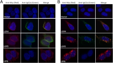
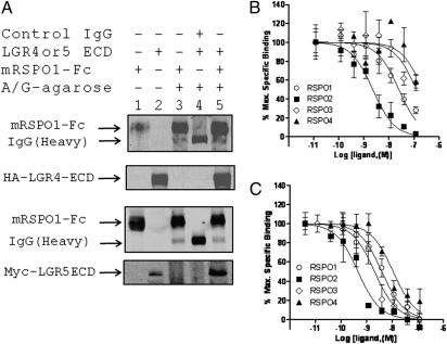
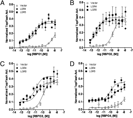
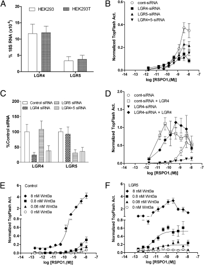
The Wnt/β-catenin signaling system plays essential roles in embryonic development and in the self-renewal and maintenance of adult stem cells. R-spondins (RSPOs) are a group of secreted proteins that enhance Wnt/β-catenin signaling and have pleiotropic functions in development and stem cell growth. LGR5, an orphan receptor of the G protein-coupled receptor (GPCR) superfamily, is specifically expressed in stem cells of the intestinal crypt and hair follicle. Knockout of LGR5 in the mouse results in neonatal lethality. LGR4, a receptor closely related to LGR5, also has essential roles in development, as its knockout leads to reduced viability and retarded growth. Overexpression of both receptors has been reported in several types of cancer. Here we demonstrate that LGR4 and LGR5 bind the R-spondins with high affinity and mediate the potentiation of Wnt/β-catenin signaling by enhancing Wnt-induced LRP6 phosphorylation. Interestingly, neither receptor is coupled to heterotrimeric G proteins or to β-arrestin when stimulated by the R-spondins, indicating a unique mechanism of action. The findings provide a basis for stem cell-specific effects of Wnt/β-catenin signaling and for the broad range of functions LGR4, LGR5, and the R-spondins have in normal and malignant growth.


Conflict of interest statement

The authors declare no conflict of interest.

Figures

Fig. 1.
Fig. 1.
Binding of mRSPO1-Fc to LGR4 and LGR5 by confocal immunofluorescence analysis. HEK293 cells stably expressing vector, Myc-LGR4, Myc-LGR5, or HA-LRP6 were incubated with mRSPO1-Fc at 4 °C (A), or at 37 °C (B). The cells were then costained with fluorescence-labeled anti-tag antibodies (Cy3–anti-Myc, Alexa594–anti-HA, both mouse IgG1 subtype) for receptor detection (red) and Alexa Fluor 488-labeled anti-IgG2a for mRSPO1-Fc detection (green). Nuclei were counterstained with ToPro-3 (blue).
Fig. 2.
Fig. 2.
Binding of RSPO1–4 to LGR4 and LGR5 by coimmunoprecipitation and competition analysis. (A) Coimmunoprecipitation of LGR4ECD and LGR5ECD with mRSPO1-Fc using protein A/G sepharose beads. Pull-down samples (lanes 3–5) were probed with anti-mouse IgG antibody, or with anti-HA (LGR4ECD), or anti-Myc (LGR5ECD) antibody for each ECD. Lanes 1 and 2 are input control. (B and C) Quantitative binding analysis using a whole-cell–based assay. HEK293 cells stably expressing Myc-LGR4 (B) or Myc-LGR5 (C) were incubated with mRSPO1-Fc plus serial dilutions of purified recombinant RSPO1–4. Maximum specific binding is defined by the difference between the data with and without mRSPO1-Fc, which is ∼50% of total binding in general. All error bars are SEM (n = 3–4).
Fig. 3.
Fig. 3.
Potentiation of Wnt/β-catenin signaling by LGR4 and LGR5 in response to RPSO1–4. HEK293T cells were transiently transfected with LGR4, LGR5, or vector, plus the β-catenin reporter plasmid Super 8× TOPFlash (firefly luciferase) and pRL-SV40 (renilla luciferase) and then stimulated with serial dilutions of purified recombinant RSPO1 (A), RSPO2 (B), RSPO3 (C), or RSPO4 (D) in the presence of Wnt3a conditioned media (CM). Firefly luciferase activity of each well was normalized to that of renilla luciferase activity of the same well. All error bars are SEM (n = 4).
Fig. 4.
Fig. 4.
Effect of LGR4 and LGR5 knockdown and Wnt3a concentration on LGR5-mediated Wnt/β-catenin signaling potentiation. (A) Expression levels of LGR4 and LGR5 in HEK293 and HEK293T cells by quantitative RT-PCR analysis. (B) Effect of LGR4 and LGR5 expression knockdown on RSPO1 response in the presence of Wnt3a CM. (C) Quantitative RT-PCR results of the expression levels of LGR4 and LGR5 in siRNA-transfected cells. (D) Rescue of RSPO1 response in LGR4-siRNA cells by cotransfecting with LGR4. (E and F) Effect of exogenous Wnt3a concentration on RSPO1 response in vector (E) and LGR5 (F) cells. All error bars are SEM (n = 4).
Fig. 5.
Fig. 5.
Effect of LGR4 and LGR5 expression knockdown and LGR5 overexpression on Wnt3a-RSPO1–induced LRP6 phosphorylation and β-catenin accumulation. (A) Effect of LGR4 and LGR5 expression knockdown on endogenous response to Wnt3a-RSPO1 in LRP6 phosphorylation and β-catenin levels. HEK293T cells were transfected with control, LGR4-, or LGR5-siRNA, or both, and 2 d later, the cells were stimulated with RSPO1 (4 nM), or Wnt3a (3 nM), or both for 3 h. Phospho-LRP6 at Ser1490 (pLRP6), total LRP6 (tLRP6), nonmembrane-associated β-catenin (membrane-bound β-catenin was removed with Con A-sepharose beads), and β-actin (loading control) were then probed by immunoblot analysis. (B) Change in pLRP6 and β-catenin levels in response to RSPO1 and Wnt3a treatment in vector and LGR5-overexpressing cells. HEK293 cells stably expressing vector or LGR5 were stimulated with RSPO1 (0, 3, and 10 ng/mL) with or without Wnt3a CM for 3 h, and probed as above. (C) Time course of LRP6 phosphorylation and changes in β-catenin levels following Wnt3a and RSPO1 treatment. The cells were stimulated with RSPO1 (100 ng/mL) and Wnt3a CM for 0–6 h and probed as above.

References

    1. Barker N, et al. Identification of stem cells in small intestine and colon by marker gene Lgr5. Nature. 2007;449:1003–1007. - PubMed
    1. Jaks V, et al. Lgr5 marks cycling, yet long-lived, hair follicle stem cells. Nat Genet. 2008;40:1291–1299. - PubMed
    1. McDonald T, et al. Identification and cloning of an orphan G protein-coupled receptor of the glycoprotein hormone receptor subfamily. Biochem Biophys Res Commun. 1998;247:266–270. - PubMed
    1. Hsu SY, Liang SG, Hsueh AJ. Characterization of two LGR genes homologous to gonadotropin and thyrotropin receptors with extracellular leucine-rich repeats and a G protein-coupled, seven-transmembrane region. Mol Endocrinol. 1998;12:1830–1845. - PubMed
    1. Hermey G, Methner A, Schaller HC, Hermans-Borgmeyer I. Identification of a novel seven-transmembrane receptor with homology to glycoprotein receptors and its expression in the adult and developing mouse. Biochem Biophys Res Commun. 1999;254:273–279. - PubMed

Publication types

MeSH terms