. 2011 Aug;156(4):2172-83.
doi: 10.1104/pp.111.181180. Epub 2011 Jun 21.

Model-based analysis of Arabidopsis leaf epidermal cells reveals distinct division and expansion patterns for pavement and guard cells

Affiliations

Model-based analysis of Arabidopsis leaf epidermal cells reveals distinct division and expansion patterns for pavement and guard cells

Leila Kheibarshekan Asl et al. Plant Physiol. 2011 Aug.

Abstract

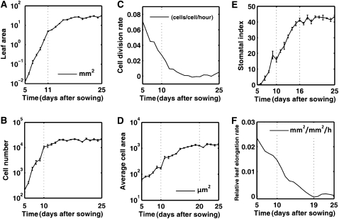
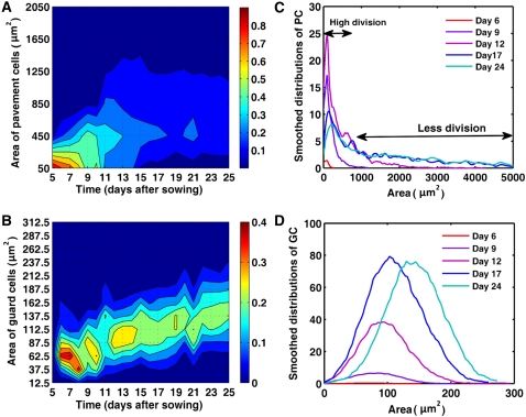
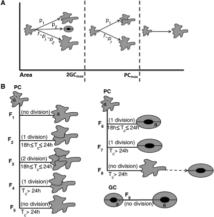
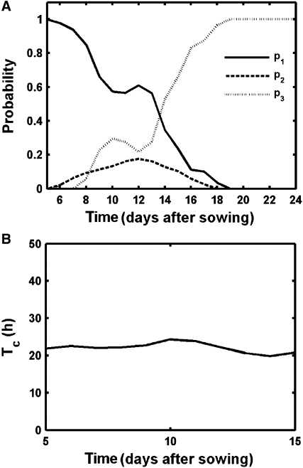
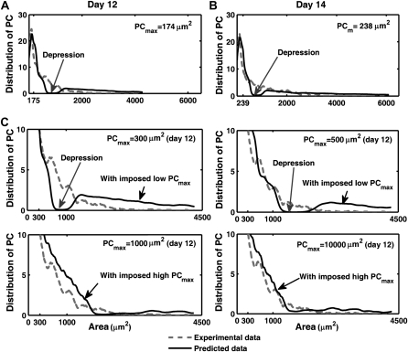
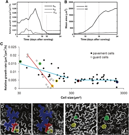
To efficiently capture sunlight for photosynthesis, leaves typically develop into a flat and thin structure. This development is driven by cell division and expansion, but the individual contribution of these processes is currently unknown, mainly because of the experimental difficulties to disentangle them in a developing organ, due to their tight interconnection. To circumvent this problem, we built a mathematic model that describes the possible division patterns and expansion rates for individual epidermal cells. This model was used to fit experimental data on cell numbers and sizes obtained over time intervals of 1 d throughout the development of the first leaf pair of Arabidopsis (Arabidopsis thaliana). The parameters were obtained by a derivative-free optimization method that minimizes the differences between the predicted and experimentally observed cell size distributions. The model allowed us to calculate probabilities for a cell to divide into guard or pavement cells, the maximum size at which it can divide, and its average cell division and expansion rates at each point during the leaf developmental process. Surprisingly, average cell cycle duration remained constant throughout leaf development, whereas no evidence for a maximum cell size threshold for cell division of pavement cells was found. Furthermore, the model predicted that neighboring cells of different sizes within the epidermis expand at distinctly different relative rates, which could be verified by direct observations. We conclude that cell division seems to occur independently from the status of cell expansion, whereas the cell cycle might act as a timer rather than as a size-regulated machinery.


Figures

Figure 1.
Figure 1.
Kinematic growth data for the first true leaf pair of Arabidopsis seedlings. A, Leaf area. B, Total cell number. C, Cell division rate. D, Mean cell area. E, Stomatal index. F, Relative leaf elongation rate.
Figure 2.
Figure 2.
Cell size distributions during leaf development. A and B, Size distribution of pavement (A) and guard (B) cells in the abaxial epidermis of leaves 1 and 2. Color ranges indicate the fraction of the total number of cells present within a specific size bin. The vertical axis shows bins of cell areas (bin size is 400 μm2 and 25 μm2 for pavement and guard cells, respectively). Labels on the vertical axis mark the beginning of each bin. Red and blue correspond to high and low percentage of cells, respectively. The colors in the ranges between two consecutive bins are obtained by interpolation. C and D, Distribution of the number of pavement (C) and guard (D) cells per leaf during its development.
Figure 3.
Figure 3.
Different scenarios of division and expansion of pavement and guard cells over a 1-d interval. A, Size categories of pavement cells. B, Global scheme of the possible flows for pavement and guard cells. The flows are defined based on the fates of the cells and the duration of the cell cycle. PC and GC, Pavement and guard cells, respectively.
Figure 4.
Figure 4.
Predicted cell division parameters during leaf development. A, Probabilities for alternative divisions; p1: pavement cells, p2: guard cells, p3: no division. B, Average cell cycle duration.
Figure 5.
Figure 5.
Observed and predicted pavement cell size distribution with optimized and imposed PCmax values. A and B, Comparison between the experimental and predicted pavement cell size distribution with optimized PCmax for day 12 (A) and day 14 (B). C, Comparison for different imposed values of PCmax at day 12. PC, Pavement cells.
Figure 6.
Figure 6.
Differential cell growth rates during leaf development. A, Average RGRs of pavement (gPC) and guard cells (gGC). gPC is the experimentally observed average RGR of small pavement cells from days 18 to 25. B, Experimental mean area of pavement and guard cells. C to E, Live imaging of epidermal cell growth in leaves. C, RGRs of pavement and guard cells on the abaxial epidermis of leaves 1 and 2 of 17-d-old plants over a period of 36 h. Red and blue lines indicate a logistic regression for guard and pavement cells, respectively. Colored dots represent data of individual pavement and guard cells depicted in sections D and E. D, Time-lapse observations of epidermal cell development over a 36-h interval. E, Time-lapse observation of guard cells over a 24-h interval. Scale bars = 25 μm.

References

    1. Baskin TI. (2000) On the constancy of cell division rate in the root meristem. Plant Mol Biol 43: 545–554 - PubMed
    1. Beemster GTS, Baskin TI. (1998) Analysis of cell division and elongation underlying the developmental acceleration of root growth in Arabidopsis thaliana. Plant Physiol 116: 1515–1526 - PMC - PubMed
    1. Beemster GTS, De Veylder L, Vercruysse S, West G, Rombaut D, Van Hummelen P, Galichet A, Gruissem W, Inzé D, Vuylsteke M. (2005) Genome-wide analysis of gene expression profiles associated with cell cycle transitions in growing organs of Arabidopsis. Plant Physiol 138: 734–743 - PMC - PubMed
    1. Beemster GTS, Fiorani F, Inzé D. (2003) Cell cycle: the key to plant growth control? Trends Plant Sci 8: 154–158 - PubMed
    1. Benková E, Michniewicz M, Sauer M, Teichmann T, Seifertová D, Jürgens G, Friml J. (2003) Local, efflux-dependent auxin gradients as a common module for plant organ formation. Cell 115: 591–602 - PubMed

Publication types

LinkOut - more resources