. 2011 Jun;23(6):2106-24.
doi: 10.1105/tpc.111.085464. Epub 2011 Jun 21.

Developmental analysis of a Medicago truncatula smooth leaf margin1 mutant reveals context-dependent effects on compound leaf development

Affiliations

Developmental analysis of a Medicago truncatula smooth leaf margin1 mutant reveals context-dependent effects on compound leaf development

Chuanen Zhou et al. Plant Cell. 2011 Jun.

Abstract

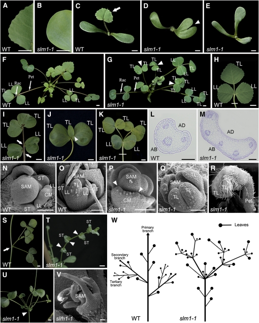
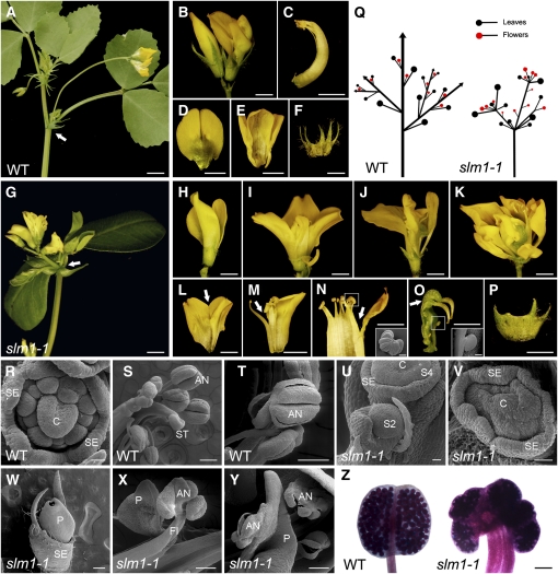
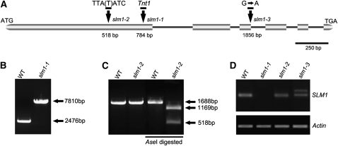
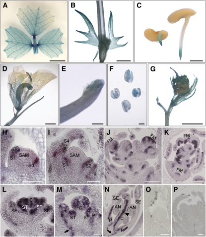
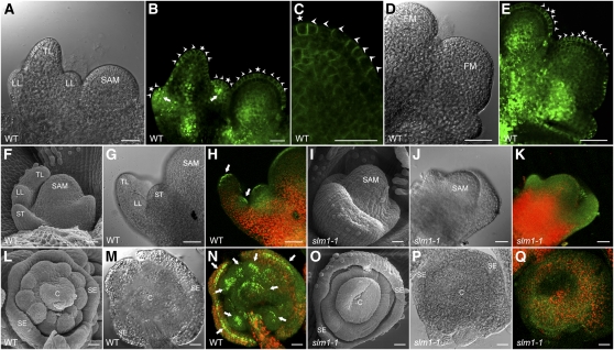
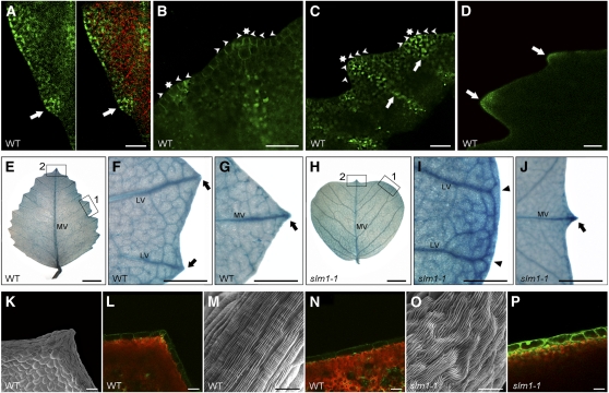
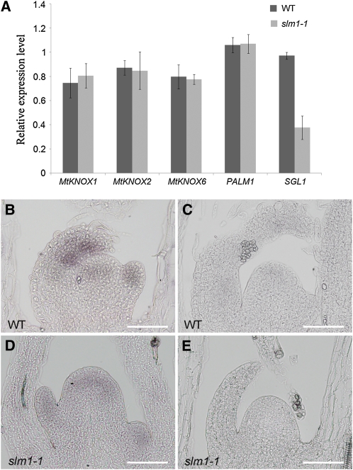
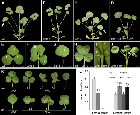
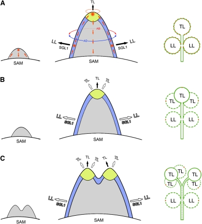
Compound leaf development requires highly regulated cell proliferation, differentiation, and expansion patterns. We identified loss-of-function alleles at the SMOOTH LEAF MARGIN1 (SLM1) locus in Medicago truncatula, a model legume species with trifoliate adult leaves. SLM1 encodes an auxin efflux carrier protein and is the ortholog of Arabidopsis thaliana PIN-FORMED1 (PIN1). Auxin distribution is impaired in the slm1 mutant, resulting in pleiotropic phenotypes in different organs. The most striking change in slm1 is the increase in the number of terminal leaflets and a simultaneous reduction in the number of lateral leaflets, accompanied by reduced expression of SINGLE LEAFLET1 (SGL1), an ortholog of LEAFY. Characterization of the mutant indicates that distinct developmental domains exist in the formation of terminal and lateral leaflets. In contrast with the pinnate compound leaves in the wild type, the slm1 sgl1 double mutant shows nonpeltately palmate leaves, suggesting that the terminal leaflet primordium in M. truncatula has a unique developmental mechanism. Further investigations on the development of leaf serrations reveal different ontogenies between distal serration and marginal serration formation as well as between serration and leaflet formation. These data suggest that regulation of the elaboration of compound leaves and serrations is context dependent and tightly correlated with the auxin/SLM1 module in M. truncatula.


Figures

Figure 1.
Figure 1.
The slm1-1 Mutant of M. truncatula Shows Developmental Defects at the Vegetative Stage. (A) and (B) Leaf margin of the wild type (WT) (A) and slm1-1 (B). (C) to (E) Four-day-old seedlings of the wild type (C) and slm1-1 ([D] and [E]). The arrow points to the first true leaf in the wild type (C). Note that the development of the first true leaf was abolished in slm1-1 ([D] and [E]). The arrowhead points to a cotyledon fusion (D). (F) and (G) Five-week-old plants of the wild type (F) and slm1-1 (G). Arrowheads point to three adult leaves of slm1-1. Two of the adult leaves have double terminal leaflets developed at the distal end of petiole, and one has three terminal leaflets (G). No lateral leaflets developed in all three marked adult leaves (G). Rac, rachis; Pet, petiole; TL, terminal leaflet; LL, lateral leaflet. (H) to (K) Adult leaves of the wild type (H) and slm1-1 ([I] to [K]). Note that three terminal leaflets developed on the petiole and no lateral leaflets were produced (J). Petiole fusion could be observed in (K). Two terminal leaflets developed on the distal end of each petiole, respectively (K). Arrows indicate asymmetric lateral leaflets on the petiole (I). Arrowhead indicates two fused terminal leaflets (J). TL, terminal leaflet; LL, lateral leaflet. (L) and (M) Transverse sections of petioles in the wild type (L) and slm1-1 (M); the sectioning regions are shown in (H) and (K) by white lines, respectively. AD, adaxial side; AB, abaxial side. (N) to (R) Scanning electron micrographs of leaf primordia in the wild type at stage 3 (N) and stage 5 (O) and in slm1-1 at stage 3 (P) and stage 5 (Q), and the developing leaf in slm1-1 at stage 9 (R). Arrowheads point out that at least two terminal leaflet primordia initiated from a common leaf primordium (P). CM, common leaf primordium; TL, terminal leaflet primordium; LL, lateral leaflet primordium; ST, stipule primordium; Pet, petiole. (S) to (V) Development of branches in the wild type (S) and slm1-1 ([T] to [V]). Arrow points to the node that bears one trifoliate and a higher-order branch in the wild type (S). Arrowheads point to nodes without branches and leaves in (T) and to the distal portion of stem with radial multiple leaves and branches in (U). Scanning electron micrograph shows the SAM with radial lateral organs in slm1-1 (V). ST, stipule. (W) A schematic illustration of branch arrangement in the wild type (left) and slm1-1 (right) at the vegetative stage. Bars = 5 mm in (A) to (K) and (S) to (U), 200 μm in (L) and (M), and 50 μm in (N) to (R) and (V).
Figure 2.
Figure 2.
The slm1-1 Mutant of M. truncatula Shows Developmental Defects at the Reproductive Stage. (A) Flower development in the wild type. Arrow indicates a node that bears two open flowers and one fully expanded trifoliate. (B) Flower phenotype in the wild type. The flowers of the wild type show bilateral symmetry along the dorsal-ventral axis. (C) to (F) Dissected floral organs of the wild type. The side view of the central carpel (C), top view of vexillum (D), alae and keel (E), and sepal (F). (G) Flower development in slm1-1. Arrow indicates that flowers and leaves develop radially at the distal portion of stem. (H) to (K) Flower phenotype in slm1-1 with mild (H), moderate ([I] and [J]), and severe (K) alterations. (L) to (P) Dissected floral organs of slm1-1. Fusions between floral organs were frequently observed; for example, the fusion between vexillums (L), between stamen and petal (M), between anthers (N), and between pistils (O). The sepal is also abnormal (P). The insets in (N) and (O) show fused anthers and exposed ovules by scanning electron microscopy, respectively. Arrows in (L) to (O) indicate the fusion of floral organs. (Q) A schematic illustration of branch arrangement of the wild type (left) and slm1-1 (right) at the reproductive stage. (R) to (T) Scanning electron microscopy analysis of floral organs in the wild type. Representative images show floral primordia at stage 6 (R), anthers and stigma (S), and dehiscing anthers (T) in a mature flower. (U) to (Y) Scanning electron microscopy analysis of floral organs in slm1-1. Representative images show floral primordia at stage 2 ([U], S2), stage 4 ([U], S4), and stage 6 (V). At the late stage of floral development, fully fused petals (W), fused filament and petal (X), and dehiscing anther (Y) were observed. (Z) Pollen staining in the wild type (left) and slm1-1 (right). C, carpel; SE, sepal; P, petal; ST, stigma; AN, anther; FI, filament; S2, stage 2; S4, stage 4; WT, wild type. Bars = 5 mm in (A) and (G), 2 mm in (B) to (F) and (H) to (P), 200 μm in (S), (T), (W) to (Y), and the insets in (N) and (O), 100 μm in (Z), and 50 μm in (R), (U), and (V).
Figure 3.
Figure 3.
Molecular Characterization of SLM1 in M. truncatula. (A) Schematic diagram of the gene structure of SLM1. The positions of the ATG start and TGA stop codons are shown. Vertical arrows mark the nucleotide changes in various slm1 alleles. Numbers indicate nucleotide positions of the site of mutations. Boxes represent exons and lines represent introns. A single base, T (thymine), was deleted in slm1-2. (B) PCR amplification of SLM1 from the wild type (WT) and slm1-1. A single insertion of the tobacco Tnt1 retrotransposon (~5.3 kb) was detected in slm1-1. (C) Transcripts of SLM1 from the wild type and slm1-2 were amplified by RT-PCR and digested by AseI, resulting in length polymorphism because of a single-base-pair deletion mutation in slm1-2. Three technical replicates were performed. (D) RT-PCR analysis of SLM1 transcripts in the wild type and slm1 alleles. Altered splicing of transcript in slm1-3 is shown. Actin was used as a loading control. Three technical replicates were performed.
Figure 4.
Figure 4.
Expression Pattern of SLM1 in M. truncatula. (A) to (G) Promoter-GUS fusion studies of SLM1 expression in transgenic M. truncatula. SLM1 promoter driven GUS is expressed in the adult leaf (A), stem and stipule (B), root tip of germinating seeds (C), flower (D), stigma (E), anther (F), and 5-d-old seedpod (G). (H) to (P) In situ hybridization analysis of SLM1 mRNA in vegetative and reproductive apices of the wild type. (H) and (I) Longitudinal sections of the SAM at stage 1 (S1; [H]) and stage 4 (S4; [I]). (J) and (K) Longitudinal section (J) and transverse section (K) of floral apices at stage 2. (L) to (N) Longitudinal sections of the floral apical meristem at stage 3 (L), stage 5 (M), and stage 7 (N). (O) and (P) The sense probe was hybridized and used as control. Arrows indicate vascular bundles. Arrowhead indicates the inside of the carpel. FM, floral meristem; SE, sepal; P, petal; C, carpel; ST, stamen; AN, anther. Bars = 5 mm in (A), 2 mm in (B) to (D) and (G), 200 μm in (E) and (F), and 50 μm in (H) to (P).
Figure 5.
Figure 5.
PIN1/SLM1-Dependent Auxin Gradients in Leaf and Floral Organ Formation in M. truncatula. (A) Leaf primordia of the wild type (WT) at stage 4. (B) and (C) Distribution of the ProPIN1:PIN1:GFP marker (green signal) in leaf primordia (B) and a close view of the localization of ProPIN1:PIN1:GFP marker in the SAM (C). Arrowheads mark the direction of PIN1 polarization. Asterisks indicate the auxin convergence points that mark the site of incipient primordium initiation. (D) Floral primordia of the wild type at stage 2. FM, floral meristem. (E) Distribution of the ProPIN1:PIN1:GFP marker in floral primordia. Arrowheads point to the direction of PIN1 polarization. Asterisks indicate the auxin convergence points. (F) to (K) Leaf primordia of the wild type ([F] to [H]) and slm1-1 ([I] to [K]). Leaf primordia harboring the auxin response marker DR5 (DR5rev:GFP) were observed by scanning electron microscopy ([F] and [I]), light-field microscopy ([G] and [J]), and confocal microscopy ([H] and [K]). Arrows point to auxin accumulation at the tip of lateral and terminal leaflet primordia. (L) to (Q) Floral primordia of wild-type ([L] to [N]) and slm1-1 ([O] to [Q]). Arrows point to auxin accumulation at the tip of floral organ primordia. TL, terminal leaflet primordium; LL, lateral leaflet primordium; ST, stipule; C, carpel; SE, sepal; WT, wild type. Bars = 25 μm.
Figure 6.
Figure 6.
Involvement of SLM1 in Leaf Margin Development in M. truncatula. (A) DR5rev:GFP expression maximum at the site of serration initiation of the leaf margin (green signal, left) and an overlay image with chlorophyll autofluorescence (red signal, right) in the wild type (WT). Arrows point to the site of serration initiation. (B) and (C) PIN1:PIN1-GFP expression during the development of leaf serrations. The localization of ProPIN1:PIN1-GFP reporter is polar at the site of serration initiation (B) and developing serrations (C). Asterisks indicate auxin flow converging at the tip of a serration. Arrowheads indicate the orientation of auxin flow predicated by PIN1/SLM1. Arrows point to the location of lateral vein formation. (D) DR5rev:GFP expression in developing leaf serrations. Arrows indicate auxin accumulation at the tip of serrations. (E) to (G) DR5:GUS expression in the fully expanded terminal leaflet of the wild type (E). Close views of marginal serration (empty box 1) and distal serration (empty box 2) are shown in (F) and (G), respectively. Arrows mark auxin accumulation at the tip of serrations. MV, midvein; LV, lateral vein. (H) to (J) DR5:GUS expression in a fully expanded terminal leaflet of slm1-1 (H). Close views of leaf margin (empty box 1) and distal serration (empty box 2) are shown in (I) and (J), respectively. Arrowheads point to lateral veins, which do not terminate at the margins. Arrow indicates auxin accumulation at the tip of the distal serration. MV, midvein; LV, lateral vein. (K) to (P) Observation of marginal cells in the wild type ([K] to [N]) and slm1-1 ([O] and [P]). Scanning electron microscopy analysis of the surface of marginal cells at the tip (K) and the side (M) of serrations in the wild type and the surface of marginal cells in slm1-1 (O). DR5rev:GFP expression is shown in the marginal cells at the same location in the wild type ([L] and [N]) and slm1-1 (P). Bar = 25 μm in (A) to (C), 5 mm in (E) and (H), 1 mm in (F), (G), (I), and (J), 150 μm in (D), 50 μm in (K), (L), (N), and (P), and 20 μm in (M) and (O).
Figure 7.
Figure 7.
Expression Analysis of Genes Related to Compound Leaf Development in M. truncatula. (A) Transcript levels of the M. truncatula KNOX1, PALM1, and SGL1 genes in the wild type (WT) and slm1-1. Transcript levels were measured by qRT-PCR using leaf meristems from 6-week-old plants. Means ± se are shown (n = 3). (B) to (E) In situ hybridization and expression patterns of SGL1 in leaf primordia of the wild type (B) and slm1-1 (D). SGL1 sense probes were used as a negative control in the wild type (C) and slm1-1 (E). Bars = 50 μm.
Figure 8.
Figure 8.
SLM1 and SGL1 Regulate Compound Leaf Development in M. truncatula. (A) to (D) Four-week-old plants of the wild type (WT) (A), slm1-1 (B), sgl1-5 (C), and slm1-1 sgl1-5 (D). Arrows indicate the juvenile leaf in (A) and (C). Note that the juvenile leaf did not develop in (B) and (D). (E) to (J) Adult leaves of the wild type (E), slm1-1 (F), sgl1-5 (G), and slm1-1 sgl1-5 ([H] and [I]). Close view of the basal region of terminal leaflets of slm1-1 sgl1-5 (I). Note that three terminal leaflets were developed on the distal end of petiole (I). Radial multiple leaves developed at the distal portion of the stem in slm1-1 sgl1-5 (J). TL, terminal leaflet; LL, lateral leaflet. (K) Adult leaf phenotype of wild-type (top) and sgl1-5 (bottom) plants grown on MS medium supplemented with 50 μM NPA. Control plants were grown on MS medium supplemented with the same concentration of DMSO. The letters a to d indicate variations of compound leaf forms in the wild type and sgl1-5 under NPA treatment. (L) Number of lateral leaflets and terminal leaflets in the wild type and the mutants. Fifty-day-old plants were used for calculating the leaflet numbers of adult leaves. Means ± se are shown (n = 100). Bars = 5 mm in (A) to (H), (J), and (K), and 2 mm in (I).
Figure 9.
Figure 9.
A Proposed Model for Compound Leaf Development Regulated by Auxin Polarity in M. truncatula. (A) In the wild type, an incipient primordium is initiated at the flanks of the SAM (left). The convergence of epidermal auxin flow (red arrows) forms a maximum of auxin activity (asterisk) at the tip of the primordium and then the auxin is drained through the center of the primordium. During the development of the leaf primordium (middle), dorsiventral polarity (the part of lower circle with blue color: adaxial side; the part of lower circle with red color: abaxial side) is established and the pseudomeristematic region termed blastozone at the margin of the primordium is formed (blue). The auxin flow converges again to form the maxima of auxin activity (asterisk), marking the sites of incipient lateral leaflet (LL) primordia formation at the blastozone. Auxin activity maxima are also formed at the tip of the terminal zone (yellow), which gives rise to the terminal leaflet (TL) primordium. The terminal zone is probably more likely to resemble the SAM with a radial prepattern (the upper circle with orange color) than to develop dorsiventral polarity (middle). The initiation of lateral leaflet primordia is in an SGL1-dependent manner, but terminal leaflet development does not depend on SGL1. The formation of serrations on the leaf margin also correlates with auxin activity maxima (orange spots) at the tip of serrations (right). (B) and (C) A developmental model of compound leaves in the slm1 mutant. As a result of diffuse auxin distribution in the slm1 mutant, the incipient primordia are able to initiate ([B], left), but fused leaf primordia initiate in some cases ([C], left), resulting in the formation of double terminal zones ([C], middle) and fused petioles ([C], right, broken line). Compared with the wild type, fewer lateral leaflet primordia (0 to 2) develop at the blastozone in an SGL1-dependent manner (empty fonts). However, the terminal zone has the potential to develop one to three terminal leaflet primordia ([B], middle; [C], middle). In addition, the leaf margin of slm1, except the distal serration (orange spots), becomes entire due to the abolished local auxin gradient activity ([B], right; [C], right). The broken line circle in (B) and (C) represents potential leaflets in slm1.

References

    1. Alexander M.P. (1969). Differential staining of aborted and nonaborted pollen. Stain Technol. 44: 117–122 - PubMed
    1. Balla J., Kalousek P., Reinöhl V., Friml J., Procházka S. (2011). Competitive canalization of PIN-dependent auxin flow from axillary buds controls pea bud outgrowth. Plant J. 65: 571–577 - PubMed
    1. Barkoulas M., Hay A., Kougioumoutzi E., Tsiantis M. (2008). A developmental framework for dissected leaf formation in the Arabidopsis relative Cardamine hirsuta. Nat. Genet. 40: 1136–1141 - PubMed
    1. Bayer E.M., Smith R.S., Mandel T., Nakayama N., Sauer M., Prusinkiewicz P., Kuhlemeier C. (2009). Integration of transport-based models for phyllotaxis and midvein formation. Genes Dev. 23: 373–384 - PMC - PubMed
    1. Benková E., Michniewicz M., Sauer M., Teichmann T., Seifertová D., Jürgens G., Friml J. (2003). Local, efflux-dependent auxin gradients as a common module for plant organ formation. Cell 115: 591–602 - PubMed

Publication types

MeSH terms

LinkOut - more resources