. 2011 Jun 21;30(14):2853-67.
doi: 10.1038/emboj.2011.204.

Autoregulation of Parkin activity through its ubiquitin-like domain

Affiliations

Autoregulation of Parkin activity through its ubiquitin-like domain

Viduth K Chaugule et al. EMBO J. .

Abstract

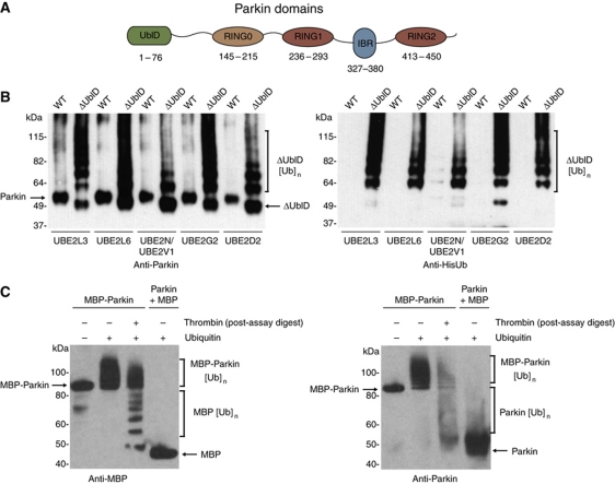
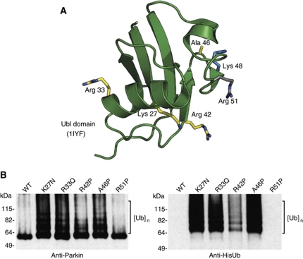
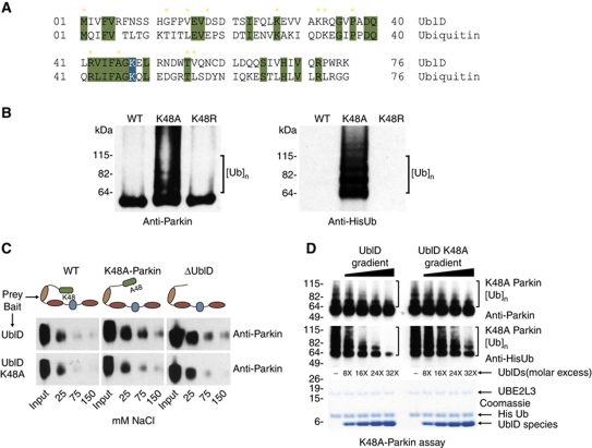
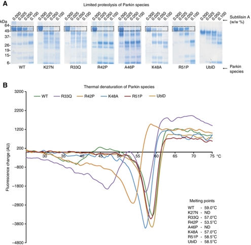
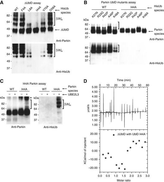
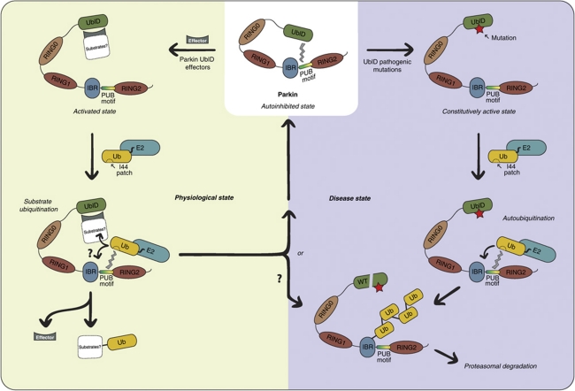
Parkin is an E3-ubiquitin ligase belonging to the RBR (RING-InBetweenRING-RING family), and is involved in the neurodegenerative disorder Parkinson's disease. Autosomal recessive juvenile Parkinsonism, which is one of the most common familial forms of the disease, is directly linked to mutations in the parkin gene. However, the molecular mechanisms of Parkin dysfunction in the disease state remain to be established. We now demonstrate that the ubiquitin-like domain of Parkin functions to inhibit its autoubiquitination. Moreover pathogenic Parkin mutations disrupt this autoinhibition, resulting in a constitutively active molecule. In addition, we show that the mechanism of autoregulation involves ubiquitin binding by a C-terminal region of Parkin. Our observations provide important molecular insights into the underlying basis of Parkinson's disease, and in the regulation of RBR E3-ligase activity.


Conflict of interest statement

The authors declare that they have no conflict of interest.

Figures

Figure 1
Figure 1
The ubiquitin-like domain of Parkin inhibits its autoubiquitination. (A) Schematic representation of Parkin molecule. The ubiquitin-like (UblD), RING0 (R0), RING (R1, R2) and in-between-RING (IBR) domains are indicated. The numbering for each domain is as per the human protein. (B) Western blot analysis of autoubiquitination assays of WT Parkin (WT) and ΔUbl-Parkin (ΔUblD) reveal inhibition of Parkin autoubiquitination by the Ubl domain. Formation of ubiquitin conjugates is detected with α-Parkin (left) and α-His-Ub (right) and indicated with a bracket. The E2 used in each experiment is indicated. (C) Western blot analysis of autoubiquitination assays of MBP-Parkin, showing competence for autoubiquitination only in the fused protein. Thrombin was added after the reaction has been stopped, leading to the cleavage of ubiquitinated MBP-Parkin. Detection of conjugates by MBP and Parkin-specific antibodies reveals that most of the ubiquitin moieties are on the MBP tag (lane 3). Lane 1 has no ubiquitin. Brackets indicate ubiquitination.
Figure 2
Figure 2
Pathogenic mutations in the Ubl domain relieve autoinhibition. (A) NMR structure of human Parkin showing the location of each of the pathogenic mutations in yellow, Lys48 in blue, and the site of the silent mutation in grey (PDB code 1iyf (Sakata et al, 2003)). (B) Western blot analysis of Parkin autoubiquitination shows that pathogenic point mutations render Parkin active for autoubiquitination while R51P does not. Ubiquitin conjugates are detected using Parkin and His antibodies and are indicated by brackets. Ube2L3 is the E2 in the assay.
Figure 3
Figure 3
Lysine 48 is required for the intramolecular interaction. (A) Sequence alignment of the Ubl domain and ubiquitin. Conserved residues are highlighted in green with K48 highlighted in blue. Yellow stars indicate pathogenic Parkin mutations. (B) Western blot analysis of Parkin autoubiquitination shows K48A renders Parkin active for autoubiquitination while K48R does not. Ubiquitin conjugates are detected using Parkin and His antibodies and are indicated by brackets. (C) Western blot analysis of full-length Parkin species pulled down by either an intact Ubl domain (top panel) or mutant K48A Ubl domain (lower panel), showing that WT Ubl domain interacts with K48A-Parkin and ΔUbl-Parkin at physiological salt concentrations, while K48A-Ubl domain does not. Input and salt concentrations are indicated. (D) Western blot analysis of K48A-Parkin autoubiquitination shows that increasing concentrations of WT Ubl domain reduces K48A-Parkin autoubiquitination to a greater extent than K48A-Ubl domain. Conjugates are detected using Parkin and His antibodies and are indicated by brackets. Excess concentrations are indicated as a ratio of Ubl species to K48A-Parkin, and a coomassie-stained gel of the input is shown beneath.
Figure 4
Figure 4
The Ubl domain interacts with ΔUbl-Parkin. (A) 600 MHz 1H–15N HSQC spectra showing the interaction between 15N-labelled Ubl domain in the absence (black contours) and presence of 0.33 (cyan), 0.67 (blue) and 1.0 (magenta) equivalents of ΔUblD. Residue assignments are indicated for the Ubl domain beside each peak. The spectra show the disappearance of many peaks in the Ubl domain spectrum as a function of ΔUblD concentration, indicative of an interaction with the ΔUblD fragment. To the right, a magnified view of part of the spectra is shown. (B) ITC curves showing binding of WT UblD (left) and K48A-UblD (right) to ΔUblD-Parkin. Dissociation constants and stoichiometry of the interaction are indicated.
Figure 5
Figure 5
WT Parkin is more stable than pathogenic mutants. (A) Proteolytic digest of Parkin species in the presence of increasing percentage of subtilisin (w/w). The boxed area indicates the degradation of all full-length species of Parkin. (B) Difference scanning fluorimetry of Parkin species. Each thermal denaturation curve is coloured according to the key, and the melting point (Tm) of each protein is noted in the figure.
Figure 6
Figure 6
Ubl-binding partners but not E2 interactions ameliorate autoinhibition. (A) Coomassie and western blot analysis of Parkin autoubiquitination shows that increasing concentrations of E2s does not overcome the autoinhibition of Parkin autoubiquitination. Parkin antibodies are used for detection, and molar excess of E2 species is indicated. (B) Pull-down analysis of Parkin and ΔUblD-Parkin with Ube2L3. A beads-only control is shown (left) and Parkin retention by the E2 is analysed by coomassie staining and western blot. (C) E2∼Ub thioester discharge assay of each Parkin species. A representative western blot of His-Ube2L3 and His-Ube2L3∼His-Ub levels upon discharge induced by indicated Parkin species. Quantitation of absolute thioester levels (3 × repeats) is graphically represented (mean±s.e.m., right). Raw data were analysed using one-way ANOVA followed by Dunnett’s multiple comparison post-test using ‘0 min’ absolute thioester level (grey) as control (***P<0.001). (D) Western blot analysis of Parkin autoubiquitination in the presence of WT and mutant UIMs from the Parkin interactor EPS15. Ubiquitin conjugates are detected using Parkin and His antibodies and are indicated by brackets. (E) Western blot analysis of Parkin autoubiquitination in the presence of WT and mutant SH3 domain from the Parkin interactor, endophilin 1A. Ubiquitin conjugates are detected using Parkin antibody and are indicated by brackets.
Figure 7
Figure 7
Mechanism of Parkin autoinhibition. (A) Western blot analysis of ΔUblD autoubiquitination with mutant ubiquitin species. Ubiquitin conjugates are detected using Parkin and His antibodies and are indicated by brackets. (B) Western blot analysis of Parkin point mutants with WT (left) and I44A (right) ubiquitin. Ubiquitin conjugates are detected using Parkin and His antibodies and are indicated by brackets. (C) Western blot analysis of I44A-Parkin autoubiquitination. Ubiquitin conjugates are detected using Parkin and His antibodies and are indicated by brackets. (D) ITC curve depicting lack of interaction between I44A-UblD and ΔUblD.
Figure 8
Figure 8
Model for Parkin activation. In the physiological state (left), Parkin is autoinhibited and activated by effectors, which may in turn bind true Parkin substrates. The C-terminal RING regions recruit E2, with an additional interaction between the thioester charged ubiquitin on E2 and the PUB motif in Parkin. This may enhance substrate ubiquitination and/or Parkin autoubiquitination. In the disease state (right), a constitutively active Parkin autoubiquitinates and is targeted for proteasomal degradation.

Comment in

  • Policing Parkin with a UblD.
    Liu F, Walters KJ. Liu F, et al. EMBO J. 2011 Jul 20;30(14):2757-8. doi: 10.1038/emboj.2011.223. EMBO J. 2011. PMID: 21772326 Free PMC article.

References

    1. Biasini E, Fioriti L, Ceglia I, Invernizzi R, Bertoli A, Chiesa R, Forloni G (2004) Proteasome inhibition and aggregation in Parkinson’s disease: a comparative study in untransfected and transfected cells. J Neurochem 88: 545–553 - PubMed
    1. Campanero MR, Flemington EK (1997) Regulation of E2F through ubiquitin-proteasome-dependent degradation: stabilization by the pRB tumor suppressor protein. Proc Natl Acad Sci USA 94: 2221–2226 - PMC - PubMed
    1. Chau V, Tobias JW, Bachmair A, Marriott D, Ecker DJ, Gonda DK, Varshavsky A (1989) A multiubiquitin chain is confined to specific lysine in a targeted short-lived protein. Science 243: 1576–1583 - PubMed
    1. Chung KK, Thomas B, Li X, Pletnikova O, Troncoso JC, Marsh L, Dawson VL, Dawson TM (2004) S-nitrosylation of parkin regulates ubiquitination and compromises parkin’s protective function. Science 304: 1328–1331 - PubMed
    1. Cooper HJ, Heath JK, Jaffray E, Hay RT, Lam TT, Marshall AG (2004) Identification of sites of ubiquitination in proteins: a fourier transform ion cyclotron resonance mass spectrometry approach. Anal Chem 76: 6982–6988 - PubMed

Publication types

MeSH terms