. 2011 Jun 21;30(14):2817-28.
doi: 10.1038/emboj.2011.194.

Drosophila Set1 is the major histone H3 lysine 4 trimethyltransferase with role in transcription

Affiliations

Drosophila Set1 is the major histone H3 lysine 4 trimethyltransferase with role in transcription

M Behfar Ardehali et al. EMBO J. .

Abstract

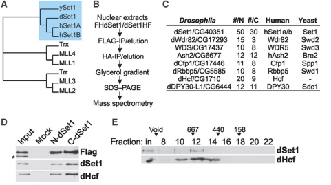
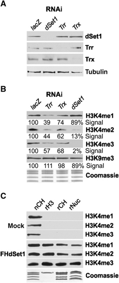
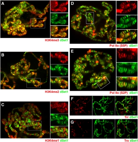
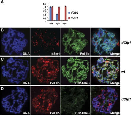
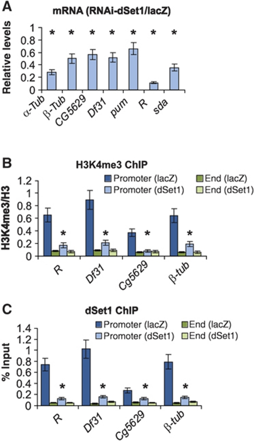
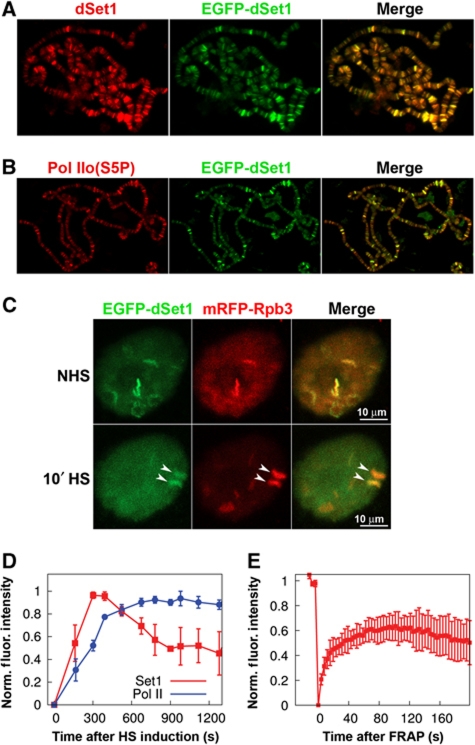
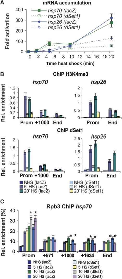
Histone H3 lysine 4 trimethylation (H3K4me3) is a major hallmark of promoter-proximal histones at transcribed genes. Here, we report that a previously uncharacterized Drosophila H3K4 methyltransferase, dSet1, and not the other putative histone H3K4 methyltransferases (Trithorax; Trithorax-related protein), is predominantly responsible for histone H3K4 trimethylation. Functional and proteomics studies reveal that dSet1 is a component of a conserved H3K4 trimethyltransferase complex and polytene staining and live cell imaging assays show widespread association of dSet1 with transcriptionally active genes. dSet1 is present at the promoter region of all tested genes, including activated Hsp70 and Hsp26 heat shock genes and is required for optimal mRNA accumulation from the tested genes. In the case of Hsp70, the mRNA production defect in dSet1 RNAi-treated cells is accompanied by retention of Pol II at promoters. Our data suggest that dSet1-dependent H3K4me3 is responsible for the generation of a chromatin structure at active promoters that ensures optimal Pol II release into productive elongation.


Conflict of interest statement

The authors declare that they have no conflict of interest.

Figures

Figure 1
Figure 1
Characterization of the Drosophila dSet1 complex. (A) Phylogenetic comparison of the Set1 and Mll1-4 homologues from yeast, flies, and humans generated with Phylip (bootstrap value 1000, 15 repeats). (B) Purification scheme for FH-dSet1 or dSet1-HF complexes. (C) Identified polypeptides from purified dSet1 complexes compared with human and yeast complexes. #N, #C: unique peptides from N- or C-terminally tagged dSet1. (D) Immunoblot analysis of purified dSet1 complexes. The asterisk marks a non-specific band. (E) Western blot analysis of nuclear extract fractionated by gel filtration. Arrowheads, calibration marker sizes in kDa.
Figure 2
Figure 2
dSet1 is required for bulk H3K4 di- and trimethylation. (A) Western blot analysis of nuclear extracts from RNAi-treated cells (top). lacZ: control from cells treated with dsRNA for E. coli lacZ. Tubulin served as loading control. (B) H3K4 methylation changes upon KD of dSet1, Trr, or Trx. Immunoblots of histone extracts from RNAi-treated cells probed with antibodies against methylated H3K4 (me1, me2, me3) or H3K9me3. Values below each lane represent the relative intensity of each band in comparison with the respective lacZ lane as quantified by ImageJ. (C) Purified dSet1 complexes methylate H3K4 in vitro. Immunoblots of KMT assays with the purified dSet1 complex (FHdSet1). Mock, purifications from mock-transfected cells. nCH, native fly histones served as positive control; rH3, recombinant H3; rCH, core histones; rNuc, nucleosomes. Coomassie, loading control.
Figure 3
Figure 3
dSet1 co-localizes with and is required for H3K4 trimethylation at transcription sites. (A) Chromosomes co-stained with antibodies against H3K4me3 (red) and dSet1 (green). (B) Chromosomes labelled with anti-H3K4me2 (red) and anti-dSet1 (green) antibodies. (C) Chromosomes stained for H3K4me1 (red) and dSet1 (green). (D) Chromosomes co-stained with antibodies against Pol IIo (S5P; red) and dSet1 (green). (E) Chromosomes double labelled with antibodies against Pol IIo (S2P; red) and dSet1 (green). (F) Chromosomes co-labelled for Trr (red) and dSet1 (green). (G) Chromosome spread stained for Trx (red) and dSet1 (green). Yellow/orange signals in the channel merges indicate co-localization. Boxed areas in (A–E) are shown in magnification to the right. Magnifications in (D) and (E) were enhanced for the red channels for better visualization of Pol IIo.
Figure 4
Figure 4
Loss of dCfp1 abolishes chromosomal association of dSet1 and H3K4me3. (A) RT/qPCR data for dCfp1 (blue) or dSet1 (red) using total RNA from salivary glands of wild-type (+/+), dCfp1 heterozygotes (+/−), or dCfp1 homozygous mutant (−/−) larvae. All values were normalized against rp49. Error bars represent the s.e.m. of three independent RNA preparations. (B) Polytene chromosomes from dCfp1 homozygous larvae co-stained with antibodies against dSet1 (red) and Pol IIo (green). (C) Polytene chromosomes of wild-type larvae co-labelled with antibodies against Pol IIo (red) and H3K4me3 (green). (D) Polytene chromosomes of dCfp1 homozygous mutants co-stained with the same antibodies as in (C). (B–D) DNA was counterstained with DAPI. Yellow-orange signals in the merged channels (right panels) indicate co-localization.
Figure 5
Figure 5
dSet1 is required for transcription and promoter-proximal H3K4 methylation. (A) RT/qPCR values from seven transcribed genes, expressed as ratio of mRNA levels from dSet1 KD versus lacZ-KD samples (=1). All values were normalized against levels of rp49. (B) ChIP/qPCR values of H3K4me3 at promoters versus ends of transcribed genes in dSet1 KD and lacZ-KD cells. The values are plotted as relative enrichment of IP chromatin compared with inputs after normalization using antibodies against H3. (C) ChIP assays evaluating the levels of dSet1 at promoters and ends of transcribed genes, plotted as percent IP versus input chromatin. Error bars represent the s.e.m. of three independent RNAi treatments (*P<0.01, t-test).
Figure 6
Figure 6
EGFP–dSet1 is rapidly recruited to the activated Hsp70 loci. Polytene chromosomes from transgenic third-instar larvae expressing EGFP–dSet1 double labelled with antibodies against (A) GFP (green) and dSet1 (red); (B) GFP (green) and Pol IIo (red); (C) laser-scanning microscopy (maximum intensity projections) shows co-localization of EGFP–dSet1 and mRFP–Rpb3 prior and after a 10-min HS in living salivary gland cells. Arrows denote the 87A and 87C HS loci that harbour multiple copies of Hsp70. (D) Recruitment of EGFP–dSet1 and mRFP–Rpb3 (Pol II) to Hsp70 loci as a function of time after HS induction (n=6, error bars represent s.e.m). (E) FRAP recovery plot showing recovery of EGFP–dSet1 signals at the Hsp70 loci. Error bars represent s.e.m. (n=8).
Figure 7
Figure 7
dSet1 is required for the maintenance of transcription from HS genes. (A) RT/qPCR data using hsp70- and hsp26-specific amplicons from dSet1 KD (dotted lines) or lacZ-KD cells (solid lines) at 0′, 2′, 5′, 10′, and 20′ at 37°C. Fold activation, signals were plotted relative to values from cells at 25°C. (B) ChIP/qPCR data from dSet1 KD cells using antibodies against H3K4me3 (middle) or dSet1 (bottom) at different HS times (0′, 5′, 20′; see legend in bottom). Amplicons at promoters (prom), about 1000 bp downstream of the TSS (+1000), and at the 3′ end (end) of the hsp70 locus were used. The hsp26 gene only spans 1010 bp. NHS, non-heat shock; HS, heat shock. (C) Pol II levels along the axis of the hsp70 gene significantly change upon the loss of dSet1 after 10′ HS (*P<0.01, t-test). Plotted are qPCR values from anti-Rpb3-ChIP using hsp70-specific amplicons normalized to signals from anti-H3CT ChIP with an amplicon targeting a non-transcribed, intergenic region. Samples from NHS, 5′, 10′, or 20′ min HS are compared. All error bars represent the s.e.m. of at least three independent RNAi treatments.

References

    1. Adelman K, Wei W, Ardehali MB, Werner J, Zhu B, Reinberg D, Lis JT (2006) Drosophila Paf1 modulates chromatin structure at actively transcribed genes. Mol Cell Biol 26: 250–260 - PMC - PubMed
    1. Allis CD, Berger SL, Cote J, Dent S, Jenuwein T, Kouzarides T, Pillus L, Reinberg D, Shi Y, Shiekhattar R, Shilatifard A, Workman J, Zhang Y (2007) New nomenclature for chromatin-modifying enzymes. Cell 131: 633–636 - PubMed
    1. Ardehali MB, Yao J, Adelman K, Fuda NJ, Petesch SJ, Webb WW, Lis JT (2009) Spt6 enhances the elongation rate of RNA polymerase II in vivo. EMBO J 28: 1067–1077 - PMC - PubMed
    1. Barski A, Cuddapah S, Cui K, Roh TY, Schones DE, Wang Z, Wei G, Chepelev I, Zhao K (2007) High-resolution profiling of histone methylations in the human genome. Cell 129: 823–837 - PubMed
    1. Boehm AK, Saunders A, Werner J, Lis JT (2003) Transcription factor and polymerase recruitment, modification, and movement on dhsp70 in vivo in the minutes following heat shock. Mol Cell Biol 23: 7628–7637 - PMC - PubMed

Publication types

MeSH terms