. 2011;6(6):e20720.
doi: 10.1371/journal.pone.0020720. Epub 2011 Jun 7.

Mammal-like organization of the avian midbrain central gray and a reappraisal of the intercollicular nucleus

Affiliations

Mammal-like organization of the avian midbrain central gray and a reappraisal of the intercollicular nucleus

Marcy A Kingsbury et al. PLoS One. 2011.

Abstract

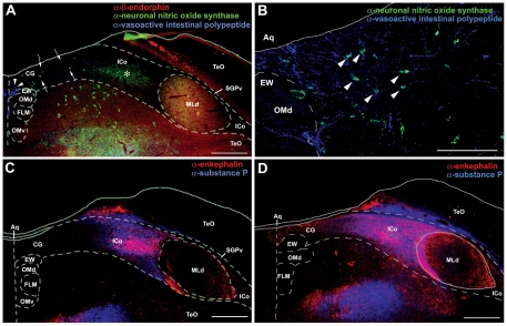
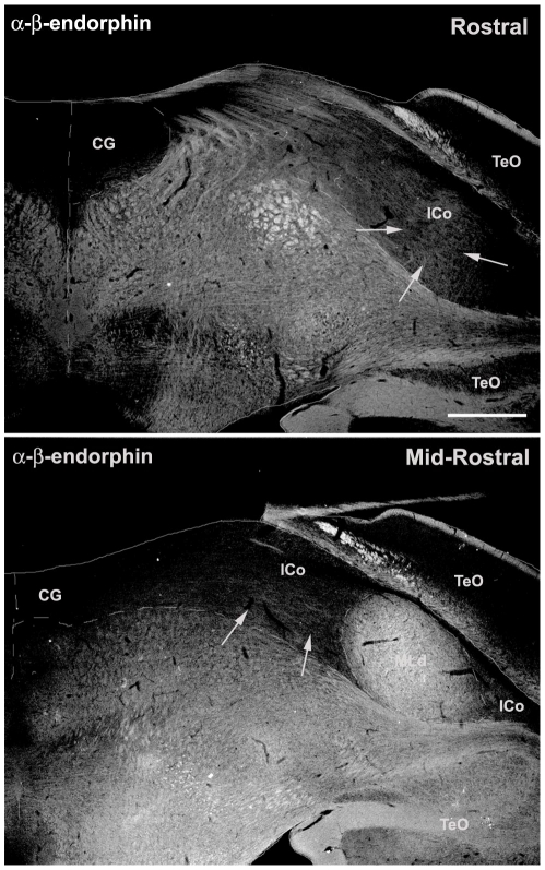
In mammals, rostrocaudal columns of the midbrain periaqueductal gray (PAG) regulate diverse behavioral and physiological functions, including sexual and fight-or-flight behavior, but homologous columns have not been identified in non-mammalian species. In contrast to mammals, in which the PAG lies ventral to the superior colliculus and surrounds the cerebral aqueduct, birds exhibit a hypertrophied tectum that is displaced laterally, and thus the midbrain central gray (CG) extends mediolaterally rather than dorsoventrally as in mammals. We therefore hypothesized that the avian CG is organized much like a folded open PAG. To address this hypothesis, we conducted immunohistochemical comparisons of the midbrains of mice and finches, as well as Fos studies of aggressive dominance, subordinance, non-social defense and sexual behavior in territorial and gregarious finch species. We obtained excellent support for our predictions based on the folded open model of the PAG and further showed that birds possess functional and anatomical zones that form longitudinal columns similar to those in mammals. However, distinguishing characteristics of the dorsal/dorsolateral PAG, such as a dense peptidergic innervation, a longitudinal column of neuronal nitric oxide synthase neurons, and aggression-induced Fos responses, do not lie within the classical avian CG, but in the laterally adjacent intercollicular nucleus (ICo), suggesting that much of the ICo is homologous to the dorsal PAG.


Conflict of interest statement

Competing Interests: The authors have declared that no competing interests exist.

Figures

Figure 1
Figure 1. Immunocytochemical comparisons of the midbrains of mice and zebra finches.
The PAG in mice (A, C, E) and the CG in finches (B, D, F) at rostral (A, B) and mid-rostral (C–F) levels of the midbrain, showing immunoreactive (-ir) cells and fibers for tyrosine hydroxylase (TH; purple), neuronal nitric oxide synthase (nNOS; red), and substance P (SP, green). Note that TH-ir cells are located ventrally along the aqueduct in mice (arrowheads in A, C, E) and medially along the aqueduct in finches (arrowheads in B, D, F) while SP-ir fibers and nNOS-ir cells are located in the lateral and dorsal columns of the PAG of mice (A, C) and in the lateral CG and ICo of finches (B, D). White arrows denote the cluster of small round nNOS-ir cells that is presumably homologous in mice and finches. TH-ir cells shown in C and D are shown at higher magnification in E and F, respectively. The schematic insets in E and F show the location of these TH-ir cells (purple dots) with respect to the aqueduct. While these neurons are located basally in both species, they are found in the ventral PAG of mice and the medial CG of finches (i.e. along red outline of aqueduct). Abbreviations for finches: Aq, aqueduct; CG, central gray; EW, Edinger-Westphal nucleus; FLM, medial longitudinal fasciculus; ICo, nucleus intercollicularis; MLd, nucleus mesencephalicus lateralis, pars dorsalis (auditory torus); nIII, oculomotor nerve; OMd/v, dorsal and ventral oculomotor nucleus; SGPv, stratum griseum periventriculare; TeO, tectum opticum. Abbreviations for mice: 3, oculomotor nucleus; bic, brachium inferior colliculus; DL, dorsolateral column of PAG; DM, dorsomedial column of PAG; L, lateral column of PAG; PAG, periaqueductal gray; PC3, parvicellular trigeminal nucleus; sc, superior colliculus; Su3, supraoculomotor central gray; Su3C, supraoculomotor cap; VL, ventrolateral column of PAG. Scale bar in A = 500 µm for A–D. Scale bar in E = 200 for E and F.
Figure 2
Figure 2. Chemoarchitecture of the avian CG/ICo.
A, Immunoreactive label for β-endorphin (β-END; red), neuronal nitric oxide synthase (nNOS; green) and vasoactive intestinal polypeptide (VIP; blue) at a mid-rostral level of the midbrain. A distinct cluster of VIP-ir cells is observed in the medial CG (arrowheads) while a discrete cluster of nNOS-ir cells is found in the lateral CG/ICo (asterisk). Diffuse immunoreactive label for VIP and nNOS is present in the dorsomedial CG (arrows, see also B). β-END-ir fibers clearly define MLd at this midbrain level, yet diffuse β-END-ir fibers can be observed in lateral ICo at both rostral and mid-rostral levels (see Figure 5). B, A higher magnification of the VIP-ir cells and fibers and nNOS-ir cells in the dorsomedial CG that were highlighted by arrows in A. Many cells within this region are co-labeled by α-VIP and α-nNOS (white arrowheads). C, D Immunoreactive label for enkephalin (ENK; red) and substance P (SP; blue) in the lateral CG/ICo at a mid-rostral (C) and caudal (D) level of the midbrain. Note that at the caudal level, ENK-ir and SP-ir extend to the midline, with ENK-ir being more prominent. See Figure 1 for abbreviations. Scale bar in A, C and D = 500 µm. Scale bar in B = 200 µm.
Figure 3
Figure 3. Immunoreactive label for substance P (SP) in the ICo surrounding MLd of a zebra finch at a mid-rostral level of the midbrain.
Note that SP-ir fibers extend through the periventricular stratum (SGPv) into the ICo lateral to MLd (arrows) and appear to define this region. See Figure 1 for abbreviations. Scale bar = 200 µm.
Figure 4
Figure 4. Immunoreactive label for neuronal nitric oxide synthase (nNOS) in the PAG of mice and CG/ICo of finches.
A, B, A distinct population of large nNOS-ir cells is present in the VL column of the PAG (A, arrowheads) and the medial CG (B, arrowhead), in addition to the cluster of smaller nNOS-ir cells in the DL column of the PAG (A, asterisk) and lateral ICo (B, asterisk). See Figure 1 for abbreviations. Scale bar = 200 µm.
Figure 5
Figure 5. β-endorphin (β-END) in the midbrain of a male zebra finch.
While β-END-ir fibers clearly define MLd at a mid-rostral level of the midbrain, diffuse β-END-ir fibers can be observed in the lateral ICo (arrows) at both rostral and mid-rostral levels. See Figure 1 for abbreviations. Scale bar = 500 µm.
Figure 6
Figure 6. An exemplar set of photomicrographs from the CG and ICo of a male zebra finch, showing the gridwork of boxes and polygons that were used for counts of Fos-ir cells at each of the three levels analyzed in zebra finches and violet-eared waxbills.
Fos-positive cells were counted in a separate Photoshop layer using the paintbrush tool (red dots). A, B Boxes and polygons used to examine Fos activation at a rostral midbrain level in the medial CG (A) and lateral CG/ICo (B). C, D Boxes and polygons used to examine Fos activation at mid-rostral (C) and caudal (D) midbrain levels. E, The triangular area of ICo lateral to MLd. This area was analyzed for each of the 3 rostrocaudal levels. Given the large individual variability in the size and shape of this area, we traced the entire triangular ICo and conducted cell counts within the outline. As with cell counts from the boxes and polygons, all Fos-ir cell counts were standardized to a unit of Fos-ir cells per 100 µm2. Based on similar response properties in contiguous sampling areas, including those that are rostrocaudally contiguous, the 36 separate sampling areas (per side) were reduced to 9 functional zones (see methods and Fig. 8A). Note that in B, C, D and E, only the left midbrain is shown, yet we analyzed both a left and right midbrain section for each animal at each rostrocaudal level. Note also that at the most rostral level analyzed, we present both the left and right medial CG in A. See Figure 1 for abbreviations. Scale bars = 250 µm.
Figure 7
Figure 7. Enhanced Fos activation in the ICo region lateral to MLd (zone 6 in Figure 8 A, D).
Greater Fos activation is observed in a dominant violet-eared waxbill following a resident intruder encounter (B) as compared to a waxbill in the control condition (A). The white arrowheads in B illustrate examples of Fos-ir cells with different levels of Fos protein in the nucleus.
Figure 8
Figure 8. Functional columns of the avian CG/ICo as delineated by Fos induction.
A, Schematic rostrocaudal representation of Fos responses in the avian CG and ICo following exposure to different social conditions (see legend). B–E, Fos-ir cell counts in male violet-eared waxbills exposed to a handled control condition (CON), nonsocial defense manipulation (pursuit by a human hand; DEF) and a resident-intruder encounter (dominant, DOM, and subordinate, SUB) within zones 1, 4, 6 and 8, respectively. F, Fos-ir cell counts in zone 9 of subordinate animals are positively correlated with the total number of aggressive behaviors received. Similar results are obtained for zones 3 and 5 (see schematic). G, Fos-ir cell counts in zone 3 of dominant animals are negatively correlated with total number of aggressive behaviors displayed. H–I, Fos-ir cell counts of male zebra finches following exposure to a handled control condition (CON), a conspecific male (MALE), a mate competition interaction (COMP; includes both courtship and aggression) and a copulatory interaction with a female (COP). J, Fos activation in zone 5 is positively correlated with the number of displacements the dominant subject directed at the competing male during mate competition. Data in panels B–E, H, I are shown as means +/− SEM. Different letters above the error bars denote significant group differences (Fisher PLSD p<0.05 following significant ANOVA). Abbreviations: AP, area pretectalis; Cb, cerebellum; CT, commissura tectalis; CP, commissura posterior; DBC, decussatio brachiorum; DM, dorsomedial nucleus; Is, nucleus interstitialis; OM, occipitomesencephalic tract; Ru, nucleus ruber; SPM, nucleus spiriformis medialis. See Figure 1 for other abbreviations.
Figure 9
Figure 9. A summary schematic illustrating the organization of various neuropeptides and neurotransmitters within the zebra finch CG and ICo across multiple rostrocaudal levels that suggest the existence of longitudinal columns based on immunohistochemistry.
The distribution of substance P (SP), vasoactive intestinal polypeptide (VIP) and neuronal nitric oxide synthase (nNOS) are shown at three rostrocaudal midbrain levels on the left while enkephalin (ENK) and tyrosine hydroxylase (TH) are shown on the right. A legend is shown at the top. The density of dots for SP-ir and x's for ENK-ir correspond to the intensity of immunoreactive fibers. Areas of the avian CG and ICo that are hypothesized to correspond to specific PAG columns in mammals are indicated in bold type at each rostrocaudal level. Note that for each neuropeptide or neuromodulator examined, each is found in a specific mediolateral position that is comparable across the different rostrocaudal levels, suggesting a longitudinal organization of neuropeptides and neuromodulators within the avian midbrain.

References

    1. Bandler R, Shipley MT. Columnar organization in the midbrain periaqueductal gray: modules for emotional expression? Trends Neurosci. 1994;17:379–389. - PubMed
    1. Behbehani MM. Functional characteristics of the midbrain periaqueductal gray. Prog Neurobiol. 1995;46:575–605. - PubMed
    1. Ruiz-Torner A, Olucha-Bordonau F, Valverde-Navarro AA, Martinez-Soriano F. The chemical architecture of the rat's periaqueductal gray based on acetylcholinesterase histochemistry: a quantitative and qualitative study. J Chem Neuroanat. 2001;21:295–312. - PubMed
    1. Sewards TV, Sewards MA. Representations of motivational drives in mesial cortex, medial thalamus, hypothalamus and midbrain. Brain Res Bull. 2003;61:25–49. - PubMed
    1. Smith GS, Savery D, Marden C, Lopez Costa JJ, Averill S, et al. Distribution of messenger RNAs encoding enkephalin, substance P, somatostatin, galanin, vasoactive intestinal polypeptide, neuropeptide Y, and calcitonin gene-related peptide in the midbrain periaqueductal grey in the rat. J Comp Neurol. 1994;350:23–40. - PubMed

Publication types