. 2011;6(6):e21175.
doi: 10.1371/journal.pone.0021175. Epub 2011 Jun 14.

LyGDI, a novel SHIP-interacting protein, is a negative regulator of FcγR-mediated phagocytosis

Affiliations

LyGDI, a novel SHIP-interacting protein, is a negative regulator of FcγR-mediated phagocytosis

Payal Mehta et al. PLoS One. 2011.

Abstract

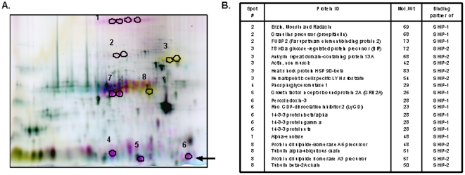
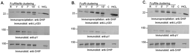
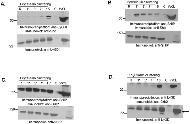
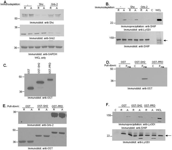
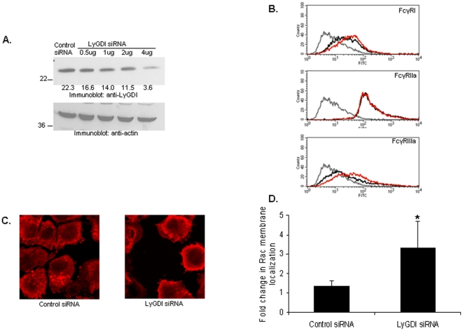
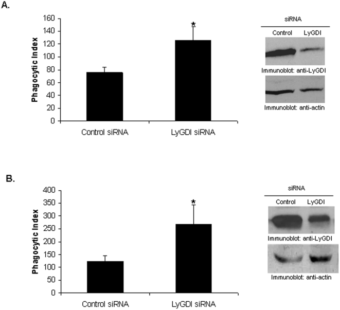
SHIP and SHIP-2 are inositol phosphatases that regulate FcγR-mediated phagocytosis through catalytic as well as non-catalytic mechanisms. In this study we have used two-dimensional fluorescence difference gel electrophoresis (DIGE) analysis to identify downstream signaling proteins that uniquely associate with SHIP or SHIP-2 upon FcγR clustering in human monocytes. We identified LyGDI as a binding partner of SHIP, associating inducibly with the SHIP/Grb2/Shc complex. Immunodepletion and competition experiments with recombinant SHIP domains revealed that Grb2 and the proline-rich domain of SHIP were necessary for SHIP-LyGDI association. Functional studies in primary human monocytes showed that LyGDI sequesters Rac in the cytosol, preventing it from localizing to the membrane. Consistent with this, suppression of LyGDI expression resulted in significantly enhanced FcγR-mediated phagocytosis.


Conflict of interest statement

Competing Interests: The authors have declared that no competing interests exist.

Figures

Figure 1
Figure 1. SHIP and SHIP-2 associate with unique binding partners.
(A) DIGE was performed as described in Materials and Methods. Spots in green are control immunoprecipitations (IP), the spots in yellow are binding partners of SHIP, while spots in red are binding partners of SHIP-2. Black circles indicate the 8 spots that were cut out and subjected to in-gel trypsin digest followed by LC-MS/MS and protein identification using a database search. The black arrow shows the spot corresponding to LyGDI. (B) This table summarizes the binding partners of SHIP or SHIP-2 that were identified.
Figure 2
Figure 2. LyGDI is a novel binding partner of SHIP.
(A) THP-1 cells were stimulated for the indicated time points using human FcγRIIa/IIb (CD32) antibody followed by goat F(ab')2 anti-mouse IgG. SHIP was immunoprecipitated from resting ‘R’ and activated ‘A’ cells and analyzed by Western blotting (IB) with anti-LyGDI (upper panel). Control IPs were performed using normal mouse IgG. The same membrane was reprobed with anti-phosphotyrosine (pY) (middle panel) and anti-SHIP (lower panel). Similar co-immunoprecipitation assays were performed from cells stimulated using (B) F(ab')2 fragments of FcγRI antibody 32.2 and goat F(ab')2 anti-mouse IgG secondary antibody, and (C) F(ab')2 fragments of FcγRIIa antibody IV.3 and goat F(ab')2 anti-mouse IgG secondary antibody.
Figure 3
Figure 3. Shc and Grb2 are present in SHIP-LyGDI complexes upon FcγR clustering.
THP-1 cells were stimulated for different time points using anti-CD32 followed by goat anti-mouse F(ab')2 IgG secondary antibody. (A) LyGDI was immunoprecipitated from resting and activated cells and analyzed by Western blotting with anti-Shc (upper panel). The same membrane was reprobed with anti-LyGDI to ensure equivalent protein loading (lower panel). Control IPs were done using normal goat IgG. (B) SHIP was immunoprecipitated from resting and activated cells and analyzed by Western blotting with anti-Shc (upper panel). The same membrane was reprobed with anti-SHIP to ensure equivalent protein loading (lower panel). Control IPs were done using normal mouse IgG. (C) SHIP was immunoprecipitated from resting and activated cells and analyzed by Western blotting with anti-Grb2 (upper panel). The same membrane was reprobed with anti-SHIP to ensure equivalent protein loading (lower panel). Control IPs were done using normal mouse IgG. (D) LyGDI was immunoprecipitated from resting and activated cells and analyzed by Western blotting with anti-Grb2 (upper panel). The same membrane was reprobed with anti-LyGDI to ensure equivalent protein loading (lower panel). Control IPs were done using normal goat IgG. All co-IP results are representative of at least three independent experiments.
Figure 4
Figure 4. Grb2 is necessary for FcγR-induced SHIP-LyGDI association.
THP-1 cells were stimulated for 15 minutes using CD32 antibody followed by goat anti-mouse IgG secondary antibody. Lysates were made and immunodepleted for Shc or Grb2. (A) Non-depleted, Shc-depleted and Grb2-depleted lysates from resting and activated cells were analyzed by Western blotting with anti-Shc (upper panel) and anti-Grb2 (middle panel). The same membrane was reprobed with anti-GAPDH (lower panel) to ensure equivalent protein loading. (B) SHIP was immunoprecipitated from resting and activated immunodepleted lysates and analyzed by Western blotting with LyGDI antibody (upper panel). The same membrane was reprobed with anti-SHIP to ensure equivalent protein loading (lower panel). Control IPs were done using normal mouse IgG. (C) 0.5 µg of purified GST, SHIP-GST-SH2 and SHIP-GST-PRD fusion proteins were analyzed by Western blotting with anti-GST antibody. (D) N-terminal biotinylated control peptide ‘C’ or phosphorylated peptide containing to the FcγRIIb ITIM sequence (PITIM) immobilized on NeutrAvidin beads were used to pulldown SHIP-GST-SH2, SHIP-GST-PRD or GST alone from solution. Bead-bound proteins were analyzed by Western blotting with GST antibody. (E) GST, SHIP-GST-SH2 or SHIP-GST-PRD were added to resting ‘R’ or FcγRIIa/IIb-stimulated ‘A’ THP-1 lysates and GST pulldowns were performed by adding glutathione agarose beads. Bead-bound proteins were analyzed by Western blotting with anti-Grb2 (upper panel). The same membrane was reprobed with anti-GST (lower panel). (F) GST, SHIP-GST-SH2 or SHIP-GST-PRD were added to resting ‘R’ or FcγRIIa/IIb-stimulated ‘A’ THP-1 lysates and LyGDI was immunoprecipiated. The immunoprecipitates were analyzed by Western blotting for SHIP (upper panel). The same membrane was reprobed with anti-LyGDI antibody (lower panel).
Figure 5
Figure 5. LyGDI influences Rac membrane localizaation.
(A) 1×10⁷ PBM were transfected with either scrambled or LyGDI siRNA. Western blotting was done to measure LyGDI after 48 hours (upper panel). The membrane was reprobed with anti-actin to ensure equivalent loading (lower panel). Numbers in the upper panel are mean optical density (arbitrary units) normalized to actin. (B) Control and LyGDI siRNA-transfected samples were tested for FcγR expression by flow cytometery analysis. Overlay histograms show FcγR staining in secondary-only antibody controls (gray), control siRNA-transfected (black) and si-LyGDI-transfected (red). (C) Control and LyGDI siRNA-transfected PBMs were stained with mouse anti-Rac followed by Alexa flour 555 conjugated to goat anti-mouse IgG, F(ab')2 fragment. (D) The fold change in Rac membrane localization in control versus LyGDI siRNA-transfected PBMs was quantified as described in Materials and Methods.
Figure 6
Figure 6. LyGDI is a negative regulator of FcγR-mediated phagocytosis.
Control and LyGDI siRNA-transfected THP-1 (A) or PBM (B) were incubated with fluoresceinated, IgG-coated sheep red blood cells (SRBC) at 37°C for 40 minutes. Phagocytosis was measured by counting the total number of SRBC ingested by 100 transfectants and is plotted as the phagocytic index. Three independent experiments were performed and scored in a blinded fashion. Data were analyzed by Student's t-test (* denotes p<0.05).

References

    1. Aderem A, Underhill DM. Mechanisms of phagocytosis in macrophages. Annu Rev Immunol. 1999;17:593–623. - PubMed
    1. Cox D, Dale BM, Kashiwada M, Helgason CD, Greenberg S. A regulatory role for Src homology 2 domain-containing inositol 5′-phosphatase (SHIP) in phagocytosis mediated by Fc gamma receptors and complement receptor 3 (alpha(M)beta(2); CD11b/CD18). J Exp Med. 2001;193:61–71. - PMC - PubMed
    1. Pengal RA, Ganesan LP, Fang H, Marsh CB, Anderson CL, et al. SHIP-2 inositol phosphatase is inducibly expressed in human monocytes and serves to regulate Fcgamma receptor-mediated signaling. J Biol Chem. 2003;278:22657–22663. - PubMed
    1. Hejna JA, Saito H, Merkens L Tittle TV, Jakobs PM, et al. Cloning and characterization of a human cDNA (INPPL1) sharing homology with inositol polyphosphate phosphatases. Genomics. 1995;29:285–287. - PubMed
    1. Erneux C, Govaerts C, Communi D, Pesesse X. The diversity and possible functions of the inositol polyphosphate 5-phosphatases. Biochim Biophys Acta. 1998;1436:185–199. - PubMed

Publication types

MeSH terms

Substances