. 2011;6(6):e20757.
doi: 10.1371/journal.pone.0020757. Epub 2011 Jun 13.

Biochemical and functional characterization of the interaction between liprin-α1 and GIT1: implications for the regulation of cell motility

Affiliations

Biochemical and functional characterization of the interaction between liprin-α1 and GIT1: implications for the regulation of cell motility

Claudia Asperti et al. PLoS One. 2011.

Abstract

We have previously identified the scaffold protein liprin-α1 as an important regulator of integrin-mediated cell motility and tumor cell invasion. Liprin-α1 may interact with different proteins, and the functional significance of these interactions in the regulation of cell motility is poorly known. Here we have addressed the involvement of the liprin-α1 partner GIT1 in liprin-α1-mediated effects on cell spreading and migration. GIT1 depletion inhibited spreading by affecting the lamellipodia, and prevented liprin-α1-enhanced spreading. Conversely inhibition of the formation of the liprin-α1-GIT complex by expression of liprin-ΔCC3 could still enhance spreading, although to a lesser extent compared to full length liprin-α1. No cumulative effects were observed after depletion of both liprin-α1 and GIT1, suggesting that the two proteins belong to the same signaling network in the regulation of cell spreading. Our data suggest that liprin-α1 may compete with paxillin for binding to GIT1, while binding of βPIX to GIT1 was unaffected by the presence of liprin-α1. Interestingly, GIT and liprin-α1 reciprocally regulated their subcellular localization, since liprin-α1 overexpression, but not the GIT binding-defective liprin-ΔCC3 mutant, affected the localization of endogenous GIT at peripheral and mature central focal adhesions, while the expression of a truncated, active form of GIT1 enhanced the localization of endogenous liprin-α1 at the edge of spreading cells. Moreover, GIT1 was required for liprin-α1-enhanced haptotatic migration, although the direct interaction between liprin-α1 and GIT1 was not needed. Our findings show that the functional interaction between liprin-α1 and GIT1 cooperate in the regulation of integrin-dependent cell spreading and motility on extracellular matrix. These findings and the possible competition of liprin-α1 with paxillin for binding to GIT1 suggest that alternative binding of GIT1 to either liprin-α1 or paxillin plays distinct roles in different phases of the protrusive activity in the cell.


Conflict of interest statement

Competing Interests: The authors have declared that no competing interests exist.

Figures

Figure 1
Figure 1. Binding of liprin-α1 to GIT1-C2 prevents binding of paxillin to GIT1-C2.
(A) Lysates were prepared from COS7 cells transfected with either HA-GIT1-C2 (C2) or co-transfected with HA-GIT1-C2 and FLAG-liprin-α1 (C2+Lip). Aliquots of the lysates were used for immunoprecipitation with anti-paxillin antibodies (IP anti-paxillin, 400 µg of protein per IP). Filters with immunoprecipitates (a), and with 100 µg of both lysates (Lys) and unbound fractions after IP (Ub) (b) were cut and immunoblotted with anti-Flag to detect Flag-liprin-α1 (upper filters, only one of the duplicated immunoprecipitations is shown); since GIT1-C2 and paxillin migrate at similar positions on gels, the lower parts of the filters from the duplicated immunoprecipitations were used as follows: one set of filters (a+b) was incubated with anti-HA to detect HA-GIT1-C2 (middle blots), and one set was incubated with anti-paxillin to detect endogenous paxillin (lower blots). Paxillin was absent from the unbound fractions after immunoprecipitation (Ub). (c) The unbound fraction (300 µg) after immunoprecipitation with anti-paxillin from the lysate of cells co-transfected with HA-GIT1-C2 and FLAG-liprin-α1 [Ub(C2+Lip)], was re-immunoprecipitated with anti-liprin antibody, to reveal the presence of the liprin-α1/GIT1-C2 complex in the lysate. (B) Binding of liprin-α1 to GIT1-C2 does not prevent binding of βPIX to GIT1-C2. Identification of a ternary complex among liprin-α1, βPIX and GIT1-C2. COS7 cells co-transfected to express the indicated combinations of HA-GIT1-C2, HA-βPIX, and FLAG-liprin-α1 were immunoprecipitated with anti-FLAG antibodies (top blots on the left). Aliquots of the unbound fraction after the first round of immunoprecipitations were re-immunoprecipitated with anti-βPIX antibodies (top blots on the right). Filters including immunoprecipitations (IP), lysates (Lys), and unbound fractions after the second round of immunoprecipitations (Ub) were cut and blotted as indicated (lower blots). (C) Liprin-α1 does not interfere with the interaction of βPIX with GIT-C2. COS7 cells co-transfected to express the indicated combinations of HA-GIT1-C2, HA-βPIX, and FLAG-liprin-α1 were immunoprecipitated with anti-βPIX antibodies. Filters including aliquots of lysates and the immunoprecipitations (IP) were cut and blotted as indicated. (D) A COS7 cell lysate (1 mg protein) was immunoprecipitated with anti-βPIX antibodies. Immunoprecipitate (IP) and equal amounts (100 µg) of lysate (Lys) and unbound fraction (Ub) were blotted with anti-GIT (mAb PKL, recognizing both GIT1 and GIT2 proteins, on the left; or anti-GIT2-specific pAb, on the right), βPIX, or anti-liprin-α1 antibodies. Blot with anti-GIT antibody was performed after stripping the filter incubated for βPIX. (E) binding of βPIX to full length GIT1 does not enhance the binding of liprin-α1 to GIT1. COS7 cells were co-transfected with FLAG-liprin-α1 and FLAG-GIT1, or with FLAG-liprin-α1 and FLAG-GIT1 and HA-βPIX. 200 µg of each lysate were immunoprecipitated with anti-GIT1 antiserum. Lysates (Lys, 50 µg), unbound fractions (Ub, 50 µg) and immunoprecipitates were blotted and incubated with antibodies specific for the indicated proteins. Overexpression of βPix did not increase the interaction of liprin-α1 with GIT1. (F) Model for the regulated interaction of GIT1 with paxillin and liprin-α1. Either ligand binds poorly to full length GIT1. We hypothesize that activation of GIT1 by so far unknown mechanisms is required for the formation of either GIT1/paxillin or GIT1/liprin-α1 complexes.
Figure 2
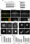
Figure 2. GIT1 and LAR depletion inhibit cell spreading and prevent enhanced spreading by liprin-α1 overexpression.
(A) Specific and control (Luc = luciferase) siRNA duplexes were used to downregulate the expression of endogenous GIT1, GIT2, liprin-α1 and LAR in COS7 cells. Cells were lysed 2 days after transfection with siRNAs. After SDS-PAGE and blotting of 50 µg of each lysate, filters were incubated with antibodies for the indicated proteins. For each specific siRNA, we could only detect the downregulation of the specific target proteins with respect to the other endogenous proteins tested as controls. For GIT1 and GIT2, a monoclonal antibody recognizing both proteins was used here. (B) The signal for endogenous GIT (red) is strongly decreased at paxillin-positive (green) focal adhesions following transfection with siRNA for either GIT1 (top) or LAR (bottom) when compared to control cells (middle). Scale bar, 5 µm. (C) COS7 cells were trypsinized 2 days after co-transfection with the indicated siRNAs and βgalactosidase (βGal), and plated 1 h on FN before immunostaining. Scale bar, 20 µm. (D, E) Quantification of spreading after replating 1 h on FN of cells co-transfected for 2 days with siRNAs (D: means ±SEM; n = 100 cells per condition), or with siRNAs and plasmids for either βgalactosidase or liprin-α1 (E: means ±SEM, n = 80–90 cells per condition from 2 experiments). **P<0.01.
Figure 3
Figure 3. Liprin-α1 affects the subcellular localization of endogenous GIT.
(A) Overexpression of liprin-α1 affects the localization of endogenous GIT at peripheral FAs. COS7 cells overexpressing either FLAG-liprin-α1 or FLAG-βgalactosidase were plated for 1 h on FN and immunostained for the transfected protein and for endogenous GIT. Scale bar, 20 µm. Right panel: four-fold enlargement of the boxed field; liprin-α1 overexpression (cell with asterisk) reduces the accumulation of GIT at newly formed FAs at the edge of transfected cells (arrowheads). Scale bar, 5 µm. (B) Cells transfected with FLAG-βgalactosidase, FLAG-liprin-α1, or FLAG-liprin-ΔCC3 were plated for 1 h on FN before fixation and staining for the transfected protein and for endogenous GIT and FAK proteins. Scale bar, 20 µm. (C) High magnification of the edge of transfected cells showing that endogenous GIT overlaps well with FAK at peripheral FAs of FLAG-liprin-ΔCC3 transfected cells, while poor overlap between endogenous GIT and FAK is seen at peripheral FAs of FLAG-liprin-α1 expressing cells. Scale bar, 10 µm. Panels on the right are 3-fold enlargements of the areas indicated by arrowheads in the corresponding images on the left.
Figure 4
Figure 4. Expression of GIT1-C affects cell morphology and the distribution of endogenous liprin-α1.
(A) COS7 cells transfected for one day with either FLAG-GIT1, FLAG-GIT1-C, or FLAG-βGalactosidase were re-plated for 1 h on FN. Immunofluorescence for the transfected proteins (FLAG), paxillin, and phalloidin staining for F-actin. Scale bar, 20 µm. Below, 3-fold enlargements of areas from cells stained for paxillin (arrowheads in the corresponding cells above) are shown. (B) Expression of GIT1-C induces a significant increase of cell spreading on FN. Bars are means ± SEM (n = 116–121 cells per condition); *P<0.05. (C) Cells transfected with either FLAG-βGalactosidase or FLAG-GIT1-C were used for triple immunofluorescence staining with antibodies for endogenous liprin-α1, paxillin, and transfected proteins (FLAG): endogenous liprin-α1 accumulates at the edge of GIT1-C-transfected cells. Scale bar, 5 µm.
Figure 5
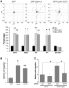
Figure 5. GIT1 is required for liprin-α1-enhanced COS7 cell migration.
(A) Transfected cells were replated on 10 µg/ml FN for 50 min to allow spreading, and then monitored for motility for 2.5 h by taking one frame every 5 min. The upper panels show cell tracks from cells transfected with the indicated constructs. The lower panel shows the quantification (mean values ±SEM) of different parameters of random migration including cell tracks (path), Euclidean distance (displ.), path rate (Vp), Euclidean rate (Vd) and persistence of migration (persist = path/displ.). N = 18–20 cells per experimental condition; *P<0.05. (B) Transwell migration assays with cells transfected with GFP, GFP-liprin-α1, or GFP-liprin-ΔCC3. Bars are normalized means ± SEM (n = 4); *P<0.05; **P<0.01. (C) Transwell migration assays with cells cotransfected with the indicated combinations of siRNAs and plasmids. Bars are normalized means ± SEM (n = 4); *P<0.05.

References

    1. Le Clainche C, Carlier MF. Regulation of actin assembly associated with protrusion and adhesion in cell migration. Physiol Rev. 2008;88:489–513. - PubMed
    1. Hoefen RJ, Berk BC. The multifunctional GIT family of proteins. J Cell Sci. 2006;119:1469–1475. - PubMed
    1. Manser E, Loo TH, Koh CG, Zhao ZS, Chen XQ, et al. PAK kinases are directly coupled to the PIX family of nucleotide exchange factors. Mol Cell. 1998;1:183–192. - PubMed
    1. Zhao ZS, Manser E, Loo TH, Lim L. Coupling of PAK-interacting exchange factor PIX to GIT1 promotes focal complex disassembly. Mol Cell Biol. 2000;20:6354–6363. - PMC - PubMed
    1. Schlenker O, Rittinger K. Structures of dimeric GIT1 and trimeric beta-PIX and implications for GIT-PIX complex assembly. J Mol Biol. 2009;386:280–289. - PubMed

Publication types

Substances