Review
. 2012 Aug;27(8):1233-47.
doi: 10.1007/s00467-011-1938-2. Epub 2011 Jun 22.

Investigating mechanisms of chronic kidney disease in mouse models

Affiliations
Review

Investigating mechanisms of chronic kidney disease in mouse models

Allison A Eddy et al. Pediatr Nephrol. 2012 Aug.

Abstract

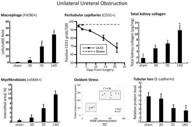
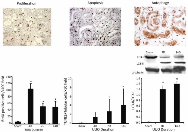
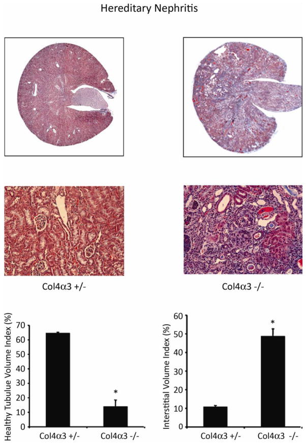
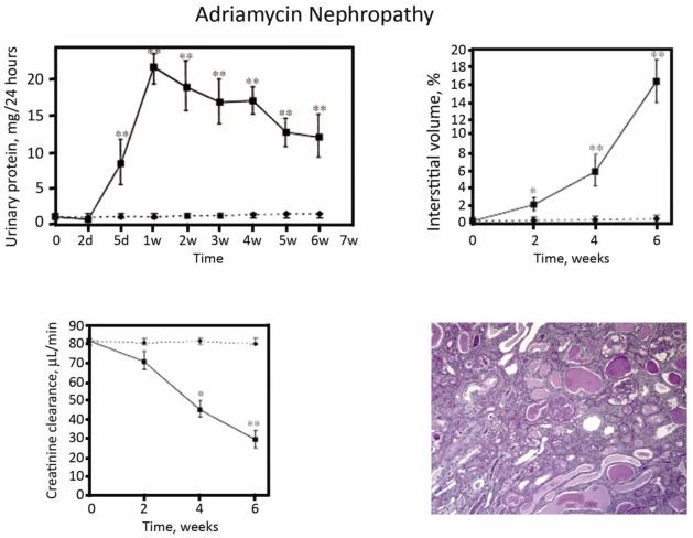
Animal models of chronic kidney disease (CKD) are important experimental tools that are used to investigate novel mechanistic pathways and to validate potential new therapeutic interventions prior to pre-clinical testing in humans. Over the past several years, mouse CKD models have been extensively used for these purposes. Despite significant limitations, the model of unilateral ureteral obstruction (UUO) has essentially become the high-throughput in vivo model, as it recapitulates the fundamental pathogenetic mechanisms that typify all forms of CKD in a relatively short time span. In addition, several alternative mouse models are available that can be used to validate new mechanistic paradigms and/or novel therapies. Here, we review several models-both genetic and experimentally induced-that provide investigators with an opportunity to include renal functional study end-points together with quantitative measures of fibrosis severity, something that is not possible with the UUO model.


Figures

Figure 1
Figure 1. Interstitial collagens I and III are major components of the interstitial scar
The fibrillar interstitial collagens I and III contribute to the formation of the interstitial matrix scaffold in normal kidneys (upper right photomicrograph); both are greatly expanded and contribute to interstitial scar formation in chronically damaged kidneys (lower right photomicrograph). Kidney mRNA levels (graph) provide a reasonable estimate of synthesis rates, as transcription is considered the rate-limiting step in collagen production. The examples are data from the unilateral ureteral obstruction (UUO) model in C57BL/6 mice (reproduced from [2, 3] reproduced with copyright permission).
Figure 2
Figure 2. Quantitative analysis of kidney collagen content
A global assessment of all kidney collagens is frequently performed using a biochemical assay to measure the content of hydroxyproline in kidney tissue, extrapolating using the assumption that 12.7% of collagen is composed of hydroxyproline (upper graph). Alternatively, kidney tissue can be stained with Sirius Red, which identifies mature, cross-linked collagen fibrils. Easily visualized by polarized light microscopy, interstitial staining is minimal in normal kidneys (left photomicrograph), which facilitates detection of abnormal deposits in diseased kidneys (right photomicrograph). Availability of computer software programs facilitates quantification collagen-positive tubulointerstitial areas (lower graph). The graphs are composites of the data from several of our published [, , –99] and unpublished studies in C57BL/6 mice. D, days after UUO surgery.
Figure 3
Figure 3. Interstitial fibrosis is heterogeneous
Interstitial fibrosis severity can vary widely from one area of the kidney to another, which underscores the importance of an adequate tissue sample size. Even in the relatively uniform injury induced by UUO of seven days’ duration, total kidney collagen varied by ~30% between different regions within the same kidney, as shown for a kidney that was harvested from a C57BL/6 mouse. The diagram depicts how the kidney was sagittally divided in half and separated into zones that correspond to each numbered bar in the graph (unpublished data).
Figure 4
Figure 4. Surrogate measures of tubular integrity
Functional compensation by the normal contralateral kidney and absence or urine flow in the obstructed kidney mean that traditional renal function outcome measures do not reflect the degree of chronic kidney damage in the UUO model. An alternative approach is to estimate the severity of the chronic kidney damage by quantifying the area of intact tubules. Possible approaches include direct measurement of tubular diameters histologically (upper photomicrograph illustrates one approach with the normal kidney on the left, a damaged kidney on the right, interstitial collagen staining in green and measured tubular diameters identified by the red lines); quantification of the area of E-cadherin-positive healthy tubules identified histologically (middle photomicrograph is from a day 14 UUO kidney) or by immunoblotting (middle graph), as shown for C57BL/6 kidneys 3 – 14 days (D) after UUO; or by estimating tubular areas using lectin staining of specific tubular segments (lotus tetragonolobus reacting with the brush border of proximal tubules indicated by the red staining of a normal mouse kidney, shown in the lower left photomicrograph; dolichos biforus identifies the stalk of the ureteric bud/collecting duct in a whole mount of a developing kidney in the lower right photomicrograph). Data in A, B and C right panel are reproduced from [13, 14, 16, 23] respectively, with copyright permission.
Figure 5
Figure 5. Unilateral ureteral obstruction (UUO): a fast and reproducible model
Despite some significant limitations, the UUO model reliably induces fibrosis with progressive kidney damage in all mouse strains reported to date, though the specific kinetics may vary between strains. The disease is characterized by the landmark histopathological features of chronic kidney disease that are illustrated above for the C57BL/6 strain and include: interstitial macrophage infiltration (F4/80 marker), interstitial capillary rarefaction (CD31 marker), oxidant stress markers (protein carbonyl and fatty acid-derived hydroxyoctadecadienoic [HODE] acid), interstitial myofibroblasts (αSMA marker), increased total kidney collagen and progressive decline in E-cadherin-positive tubules. These data are previously published and have been reproduced with copyright permission [14, 23] except the CD31 data (from Yamaguchi et al, unpublished). D, days after UUO surgery.
Figure 6
Figure 6. Mechanisms of tubular atrophy
Renal tubules are estimated to occupy 85–90% of the normal kidney volume and irreversible tubular loss is the sine qua non of chronic kidney disease. A key distinction between acute and chronic disease is the process of tubular self-repair by proliferation. Using the UUO model as an example, when proliferating tubular epithelia are labeled by an injection of bromodeoxyuridine (Brdu) that is incorporated into the nucleus of dividing cells that can be detected by immunostaining (left photomicrograph), their proliferative capacity has been shown to progressively decline with chronic injury. During this same time-period the number of tubular cells undergoing apoptotic death, detected by nuclear terminal deoxynucleotidyl transferase dUTP nick end labeling (TUNEL) staining increase (middle photomicrograph and bar graph) (data are from [14]). It has recently been reported that tubular epithelia also undergo a process of autophagy that can be identified by the expression of (LC3-II), detected by immunostaining (right photomicrograph) or immunoblotting (right graph). It is still not clear if autophagy is a protective or destructive response in the context of chronic kidney disease (data are reproduced from [21, 100]). Copyright permission was obtained for reproduction of these data.
Figure 7
Figure 7. Col4 α−/ − hereditary nephritis model
Mouse harboring a genetic mutation in the gene that encodes the glomerular basement membrane protein collagen IV alpha 3 (col4α3−/ −) develop hereditary nephritis with proteinuria and progressive renal failure. The tempo of the kidney disease is mouse strain-dependent. Compared to the asymptomatic heterozygous littermates (col4α3+/−) the mutant mice develop progressive interstitial fibrosis (shown in the Masson trichrome-stained photomicrographs), reduced tubular volume and interstitial volume expansion by matrix proteins. Data are from a study in 10-week old 129Sv/J mice that also harbor an asymptomatic heterozygous mutation in the 6 integrin gene that are reproduced from [27] with copyright permission.
Figure 8
Figure 8. Adriamycin nephropathy
Following a single intravenous injection of adriamycin (10 mg/kg), Balb/c mice develop nephrotic proteinuria, interstitial inflammation and interstitial fibrosis (represented as % interstitial volume) and renal failure (indicated as creatinine clearance). These figures are reproduced from Wang et al [56] with copyright permission. The photomicrograph is from taken from a mouse 4 weeks after adriamycin injection (unpublished).
Figure 9
Figure 9. Chronic folic acid-induced nephropathy
C57BL/6 mice given a single intraperitoneal injection of folic acid (250 mg/kg) develop acute kidney injury (shown by the serum BUN levels) but recovery is not complete. On day 14 BUN levels are still significantly elevated (64 ± 9 versus 28 ± 1 mg/dl at baseline). The kidneys show interstitial fibrosis based on computer-assisted analysis of the Masson trichrome (MT) stained interstitial area (middle graph and upper photomicrographs), accompanied by significant numbers of αSMA+ interstitial myofibroblasts (lower photomicrographs). Data are reproduced from [76] with copyright permission.
Figure 10
Figure 10. Interstitial fibrosis flowing severe acute kidney injury (AKI)
Acute renal ischemia (induced in C57BL/6 mice by cross-clamping the renal artery for 30 minutes), followed by reperfusion, has been extensively investigated as a model of acute kidney injury. Similar to humans, it is increasingly appreciated that renal recovery may not be complete; it may lead to chronic tubulointerstitial damage. In the study that is shown, (reproduced from [87] with copyright permission), the contralateral kidney was removed at the time of ischemia induction and fibrosis was detected by Masson trichrome staining 30 days later.

References

    1. Risdon RA, Sloper JC, de Vardener HE. Relationship between renal function and histologic changes found in renal-biopsy specimens from patients with persistent glomerulonephritis. Lancet. 1968;2:363–366. - PubMed
    1. Oda T, Jung YO, Kim H, Cai x, Lopez-Guisa J, Ikeda Y, Eddy AA. PAI-1 deficiency attenuates the fibrogenic response to ureteral obstruction. Kidney Int. 2001;60:587–596. - PubMed
    1. Jones CL, Buch S, Post M, McCulloch L, Liu E, Eddy AA. The pathogenesis of interstitial fibrosis in chronic purine aminonucleoside nephrosis. Kidney Int. 1991;40:1020–1031. - PubMed
    1. Farris AB, Adams CD, Brousaides N, Della Pelle PA, Collins AB, Moradi E, Smith RN, Grimm PC, Colvin RB. Morphometric and visual evaluation of fibrosis in renal biopsies. J Am Soc Nephrol. 2011;22:176–186. - PMC - PubMed
    1. Fogo AB, Alpers CE. Navigating the challenges of fibrosis assessment: land in sight? J Am Soc Nephrol. 2011;22:11–13. - PubMed

Publication types