. 2011 Jun 22:4:9.
doi: 10.1186/1757-2215-4-9.

Sodium arsenite and hyperthermia modulate cisplatin-DNA damage responses and enhance platinum accumulation in murine metastatic ovarian cancer xenograft after hyperthermic intraperitoneal chemotherapy (HIPEC)

Affiliations

Sodium arsenite and hyperthermia modulate cisplatin-DNA damage responses and enhance platinum accumulation in murine metastatic ovarian cancer xenograft after hyperthermic intraperitoneal chemotherapy (HIPEC)

Clarisse S Muenyi et al. J Ovarian Res. .

Abstract

Background: Epithelial ovarian cancer (EOC) is the leading cause of gynecologic cancer death in the USA. Recurrence rates are high after front-line therapy and most patients eventually die from platinum (Pt) - resistant disease. Cisplatin resistance is associated with increased nucleotide excision repair (NER), decreased mismatch repair (MMR) and decreased platinum uptake. The objective of this study is to investigate how a novel combination of sodium arsenite (NaAsO2) and hyperthermia (43°C) affect mechanisms of cisplatin resistance in ovarian cancer.

Methods: We established a murine model of metastatic EOC by intraperitoneal injection of A2780/CP70 human ovarian cancer cells into nude mice. We developed a murine hyperthermic intraperitoneal chemotherapy model to treat the mice. Mice with peritoneal metastasis were perfused for 1 h with 3 mg/kg cisplatin ± 26 mg/kg NaAsO2 at 37 or 43°C. Tumors and tissues were collected at 0 and 24 h after treatment.

Results: Western blot analysis of p53 and key NER proteins (ERCC1, XPC and XPA) and MMR protein (MSH2) suggested that cisplatin induced p53, XPC and XPA and suppressed MSH2 consistent with resistant phenotype. Hyperthermia suppressed cisplatin-induced XPC and prevented the induction of XPA by cisplatin, but it had no effect on Pt uptake or retention in tumors. NaAsO2 prevented XPC induction by cisplatin; it maintained higher levels of MSH2 in tumors and enhanced initial accumulation of Pt in tumors. Combined NaAsO2 and hyperthermia decreased cisplatin-induced XPC 24 h after perfusion, maintained higher levels of MSH2 in tumors and significantly increased initial accumulation of Pt in tumors. ERCC1 levels were generally low except for NaAsO2 co-treatment with cisplatin. Systemic Pt and arsenic accumulation for all treatment conditions were in the order: kidney > liver = spleen > heart > brain and liver > kidney = spleen > heart > brain respectively. Metal levels generally decreased in systemic tissues within 24 h after treatment.

Conclusion: NaAsO2 and/or hyperthermia have the potential to sensitize tumors to cisplatin by inhibiting NER, maintaining functional MMR and enhancing tumor platinum uptake.


Figures

Figure 1
Figure 1
Mouse with multiple small intraperitoneal tumors. A. MicroCT scan of tumors in live mouse. B. Direct visualization of tumors at necropsy of mouse. Three tumors are denoted by arrow in panels A and B.
Figure 2
Figure 2
Murine hyperthermic intraperitoneal chemotherapy model. A. Drawing of tumor bearing mouse undergoing HIPEC. Depicted are inlet (a) and outlet (b) ports and anal temperature probe (c) to monitor internal temperature of mouse during perfusion. B. Photograph showing perfusion pump (a), temperature monitor (b), flow tubes (c) and heating bath (d). Mice were perfused for 1 h at the rate of 3 mL/min with cisplatin (3 mg/kg) ± NaAsO2 (26 mg/kg) at 37 or 43°C.
Figure 3
Figure 3
Inductively Coupled Plasma Mass Spectrometry (ICP-MS) determination of platinum and arsenic in tumors. Mice were perfused for 1 h with cisplatin (CP/37; CP/43) or cisplatin + NaAsO2 (CPA/37; CPA/43) at 37 or 43°C respectively. Tumors from untreated (UT) and treated mice were harvested at 0 and 24 h after treatment. Tumors were homogenized and samples of the homogenate were analyzed for protein concentration by BCA or digested in nitric acid for ICP-MS analysis for platinum and arsenic. Data are presented as means ± SEM of ≥3 tumors each from different mice. Statistical analysis was performed using wilcoxon rank sum test. P < 0.05, N ≥ 3: # = lower than 0 h partner, ‡ = higher than CP/37 at 0 h and CP/43 at 0 h, ¶ = higher than CPA/43°C at 0 h.
Figure 4
Figure 4
DNA repair protein expression in tumors. A. Western blot determination of p53, XPC, XPA, ERCC1 and MSH2 in tumors. GAPDH is loading control. B. Densitometry analyses of (a) p53, (b) XPC, (c) XPA, (d) ERCC1 and (e) MSH2 normalized to GAPDH loading control and untreated tumors. Mice were perfused for 1 h with cisplatin (CP/37; CP/43) or cisplatin plus NaAsO2 (CPA/37; CPA/43) at 37 or 43°C respectively. Tumors from untreated (UT) mice and treated mice were harvested 0 and 24 h after treatment. Protein extracts were prepared from the tumors and 20 μg loaded per lane for SDS-PAGE. Data are presented as means ± SD of ≥5 tumors each from different mice. Statistical analysis was performed using wilcoxon rank sum test. P < 0.05, N ≥ 5. # = compared to 0 h partner, * = compared to UT.
Figure 5
Figure 5
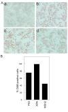
Immunocytochemical determination of p53, XPA and MSH2 expression in ovarian cancer cells. A. A2780/CP70 cells were treated for 1 h with 40 μM cisplatin. Cells were washed and incubated in drug-free media for 24 h and immunohistochemistry was performed. Representative pictures of cells at 20x magnification for secondary antibody only control (a), p53 (b), XPA (c) and MSH2 (d). B. Plot of 3,3'-diaminobenzidine (DAB)-positive cells. Data are single biological experiment performed in duplicate slides. Four different fields were counted per coverslip.
Figure 6
Figure 6
Platinum and arsenic accumulation in somatic tissues. Mice were perfused for 1 h with cisplatin (CP/37; CP/43) or cisplatin + NaAsO2 (CPA/37; CPA/43) at 37 or 43°C respectively. Tissues from untreated (UT) and treated mice were harvested at 0 and 24 h after treatment. Tissue samples were weighed and digested in nitric acid for ICP-MS analysis for platinum (A) and arsenic (B). Data are presented as means ± SD of triplicate samples each from different mice. Statistical analysis was performed using wilcoxon rank sum test. P < 0.05, N = 3. # = compared to 0 h partner.

References

    1. Jemal A, Siegel R, Ward E, Hao Y, Xu J, Thun MJ. Cancer statistics, 2009. CA Cancer J Clin. 2009;59:225–249. doi: 10.3322/caac.20006. - DOI - PubMed
    1. Armstrong DK, Bundy B, Wenzel L, Huang HQ, Baergen R, Lele S, Copeland LJ, Walker JL, Burger RA. Intraperitoneal cisplatin and paclitaxel in ovarian cancer. N Engl J Med. 2006;354:34–43. doi: 10.1056/NEJMoa052985. - DOI - PubMed
    1. Ozols RF. Treatment goals in ovarian cancer. Int J Gynecol Cancer. 2005;15(Suppl 1):3–11. - PubMed
    1. Markman M. Intraperitoneal chemotherapy in the management of malignant disease. Expert Rev Anticancer Ther. 2001;1:142–148. doi: 10.1586/14737140.1.1.142. - DOI - PubMed
    1. van d V, van d V, Zoetmulder FA, van Goethem AR, van T O, ten Bokkel Huinink WW, Beijnen JH, Bartelink H, Begg AC. Intraperitoneal cisplatin with regional hyperthermia in advanced ovarian cancer: pharmacokinetics and cisplatin-DNA adduct formation in patients and ovarian cancer cell lines. Eur J Cancer. 1998;34:148–154. doi: 10.1016/S0959-8049(97)00370-5. - DOI - PubMed