

Accuracy of linear measurement in the Galileos cone beam computed tomography under simulated clinical conditions
- PMID: 21697155
- PMCID: PMC3520257
- DOI: 10.1259/dmfr/72117593
Accuracy of linear measurement in the Galileos cone beam computed tomography under simulated clinical conditions
Abstract
Objectives: The aim of this study was to determine the geometric accuracy of cone beam CT (CBCT)-based linear measurements of bone height obtained with the Galileos CBCT (Sirona Dental Systems Inc., Bensheim, Hessen, Germany) in the presence of soft tissues.
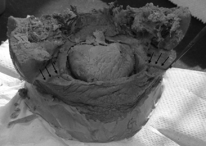
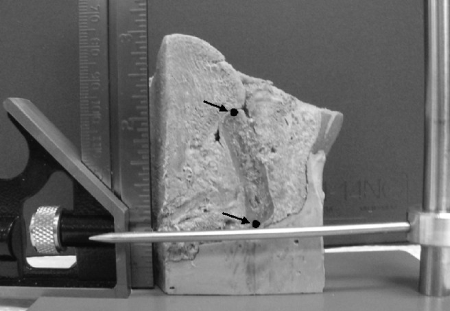
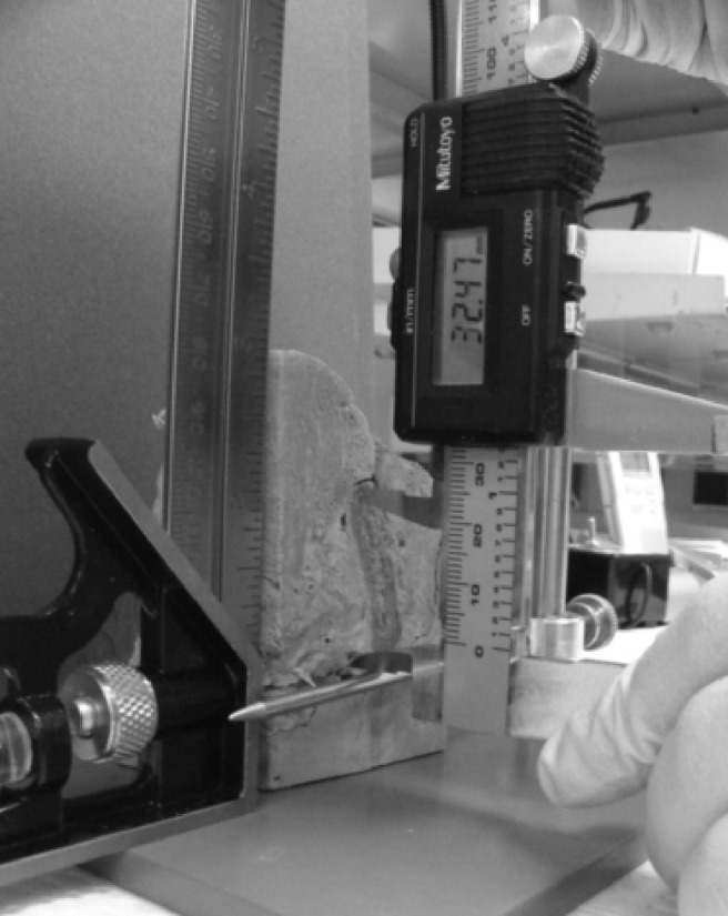
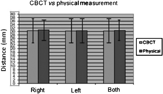
Methods: Six embalmed cadaver heads were imaged with the Galileos CBCT unit subsequent to placement of radiopaque fiduciary markers over the buccal and lingual cortical plates. Electronic linear measurements of bone height were obtained using the Sirona software. Physical measurements were obtained with digital calipers at the same location. This distance was compared on all six specimens bilaterally to determine accuracy of the image measurements.
Results: The findings showed no statistically significant difference between the imaging and physical measurements (P > 0.05) as determined by a paired sample t-test. The intraclass correlation was used to measure the intrarater reliability of repeated measures and there was no statistically significant difference between measurements performed at the same location (P > 0.05).
Conclusions: The Galileos CBCT image-based linear measurement between anatomical structures within the mandible in the presence of soft tissues is sufficiently accurate for clinical use.
Figures

References
-
- Lascala CA, Panella J, Marques MM. Analysis of accuracy of linear measurements obtained by cone beam computed tomography (CBCTNewTom). Dentomaxillofac Radiol 2004;33:291–294 - PubMed
-
- Mischkowski RA, Pulsfort R, Ritter L, Neugebauer J, Brochhagen HG, Keeve E, et al. Geometric accuracy of a newly developed cone-beam device for maxillofacial imaging. Oral Surg Oral Med Oral Pathol Oral Radiol Endod 2007;104:551–559 - PubMed
-
- Stratemann SA, Huang JC, Maki K, Miller AJ, Hatcher DC. Comparison of cone beam computed tomography imaging with physical measures. Dentomaxillofac Radiol 2008;37:80–93 - PubMed
-
- Periago DR, Scarfe WC, Moshiri M, Scheetz JP, Silveira AM, Farman AG. Linear accuracy and reliability of cone beam CT derived 3-dimensional images constructed using an orthodontic volumetric rendering program. Angle Orthodontist 2008;78:387–395 - PubMed
Publication types
MeSH terms
LinkOut - more resources
Full Text Sources
Medical
