. 2011 Jun 22;31(25):9170-8.
doi: 10.1523/JNEUROSCI.1250-11.2011.

Role of the CaMKII/NMDA receptor complex in the maintenance of synaptic strength

Affiliations

Role of the CaMKII/NMDA receptor complex in the maintenance of synaptic strength

Magdalena Sanhueza et al. J Neurosci. .

Abstract

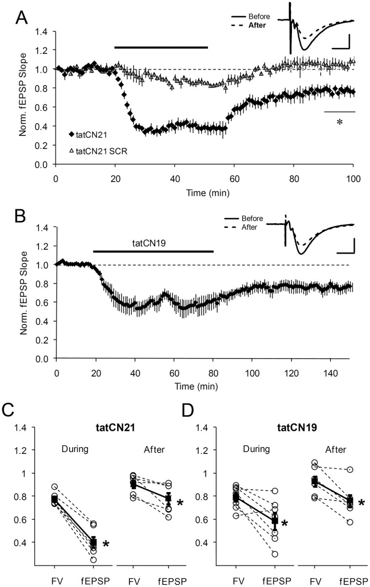
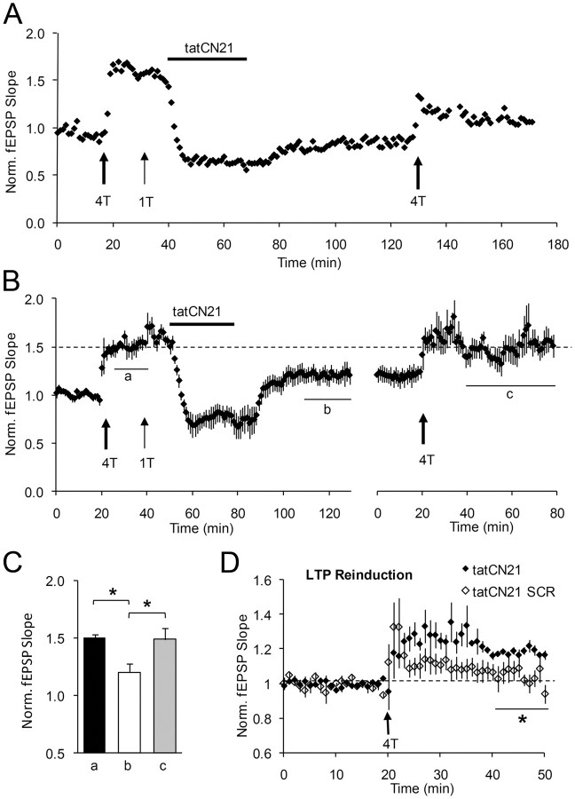
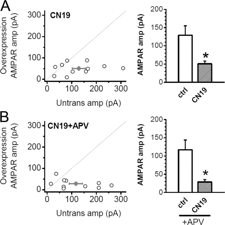
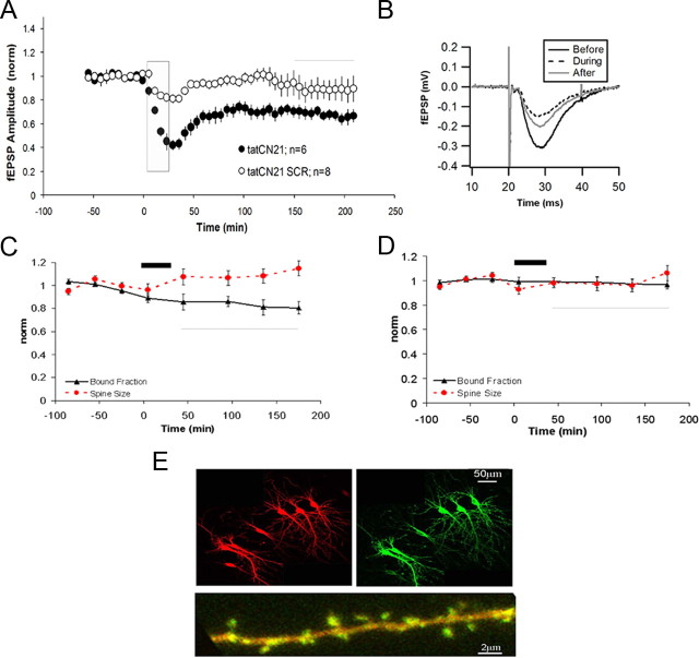
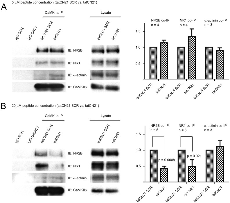
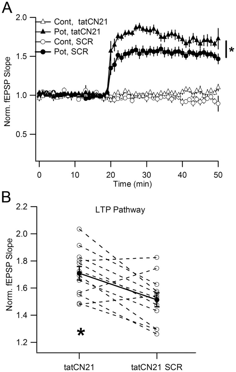
During long-term potentiation (LTP), synapses undergo stable changes in synaptic strength. The molecular memory processes that maintain strength have not been identified. One hypothesis is that the complex formed by the Ca(2+)/calmodulin-dependent protein kinase II (CaMKII) and the NMDA-type glutamate receptor (NMDAR) is a molecular memory at the synapse. To establish a molecule as a molecular memory, it must be shown that interfering with the molecule produces a persistent reversal of LTP. We used the CN class of peptides that inhibit CaMKII binding to the NR2B subunit in vitro to test this prediction in rat hippocampal slices. We found that CN peptides can reverse saturated LTP, allowing additional LTP to be induced. The peptide also produced a persistent reduction in basal transmission. We then tested whether CN compounds actually affect CaMKII binding in living cells. Application of CN peptide to slice cultures reduced the amount of CaMKII concentrated in spines, consistent with delocalization of the kinase from a binding partner in the spine. To more specifically assay the binding of CaMKII to the NMDAR, we used coimmunoprecipitation methods. We found that CN peptide decreased synaptic strength only at concentrations necessary to disrupt the CaMKII/NMDAR complex, but not at lower concentrations sufficient to inhibit CaMKII activity. Importantly, both the reduction of the complex and the reduction of synaptic strength persisted after removal of the inhibitor. These results support the hypothesis that the CaMKII/NMDAR complex has switch-like properties that are important in the maintenance of synaptic strength.


Figures

Figure 1.
Figure 1.
Persistent depression of basal transmission by bath-applied tat-fused CN peptides. A, Basal transmission was monitored before, during, and after transient application (30 min) of 20 μm tatCN21 (n = 6) or tatCN21 scrambled (SCR) peptide (n = 4). t test, *p < 0.005. Inset, Representative averaged basal fEPSP before and 50 min after drug washout. B, Same as A for 1 h application of 5 μm tatCN19 (n = 7). Inset, Averaged fEPSP before and 60 min after drug washout. C, fEPSP and fiber (FV) were reduced during peptide application; after washout, FV recovered, but there was a persistent reduction of fEPSP. Each pair represents data from a single experiment and the data were normalized relative to baseline values before drug application. Filled symbols, Average ± SEM; FV = 0.78 ± 0.02, fEPSP = 0.40 ± 0.05, during treatment; FV = 0.91 ± 0.03, fEPSP = 0.78 ± 0.05, after treatment (Wilcoxon signed test, *p < 0.05). D, Same as C, for tatCN19. FV = 0.79 ± 0.04, fEPSP = 0.58 ± 0.07, during treatment; FV = 0.93 ± 0.04, fEPSP = 0.76 ± 0.05, after treatment (*p < 0.05). Calibration: 5 ms, 0.4 mV. Error bars represent SEM in all figures.
Figure 2.
Figure 2.
TatCN21 partially reverses saturated LTP, allowing subsequent potentiation. A, Sample experiment in which saturated LTP was induced (by 4 tetani (4T), and 1 additional tetanus (1T) to show saturation). Twenty micromolar tatCN21 was next applied for 30 min, and drug was washed out for 1 h. Subsequently, LTP reinduction was tested. B, Average plot for five experiments; data were normalized to basal transmission before the first tetanization. Due to slight differences among experiments in the time for LTP reinduction (a few minutes), data were realigned with respect to tetanization time (right). C, Summary plot of mean fEPSP slope for the time intervals a–c, indicated in B; a: 1.50 ± 0.03, b: 1.20 ± 0.08, c: 1.49 ± 0.10; average ± SEM (*p < 0.01; one-way ANOVA for correlated samples, post hoc Tukey test). D, To quantify LTP reinduction, transmission was renormalized to baseline before the last series of tetani. The plot includes average data from similar experiments where SCR peptide was applied instead of tatCN21 (n = 4). After treatment with SCR peptide, tetanization induced no further potentiation, as fEPSP slope was not statistically different as compared to 20 min baseline (average ± SEM = 1.05 ± 0.05; p = 0.156), indicating that LTP saturation was unaffected. LTP reinduction after tatCN21 was 18.5 ± 1.9% (*p = 0.011; Wilcoxon rank test). The averaging interval is indicated by a continuous line.
Figure 3.
Figure 3.
CN19 decreases synaptic transmission in an NMDAR-independent way. CA1 neurons from hippocampal slice culture were transfected to overexpress GFP-CN19 for 2 d. Cell-pair recordings were performed from a transfected neuron and a nearby untransfected neuron. A, AMPAR EPSCs were significantly decreased by the overexpression of GFP-CN19 (average ± SEM: 128.7 ± 26.2 vs 50.6 ± 7.8 pA for untransfected and transfected cells, respectively; n = 11, *p = 0.013 with paired Student's t test). B, Slices were incubated in 100 μm APV after transfection for 2 d, and AMPAR EPSCs were recorded with APV removed. CN19 still significantly decreased AMPAR EPSCs after APV treatment (117.0 ± 26.9 vs 28.7 ± 6.6 pA for untransfected and transfected cells, respectively; n = 10, *p = 0.017 with paired Student's t test).
Figure 4.
Figure 4.
TatCN21 decreases synaptic transmission and bound fraction of CaMKII in dendritic spines in slice cultures. A, The graph shows a significant and persistent (for at least 3 h) decrease of fEPSP amplitude after application of 20 μm tatCN21 (black symbols), while control tatCN21 SCR peptide was ineffective. The gray bar indicates the period of drug application. B, Examples of average waveforms of fEPSP at different times during the experiment. C, Graphs showing that bound fraction of GFP-CaMKII in spines (black symbols) but not spine volume (red symbols) significantly and persistently decreases after application of tatCN21 (black bar). D, SCR peptide did not affect either bound fraction (black symbols) or spine volume (red symbols). Concentrations of both peptides were 20 μm. Thin horizontal line shows a period taken for analysis of statistical significance between control and test experiments (p < 0.001). E, Top, A representative example of CA1 neurons transfected with RFP (left) and GFP-CaMKIIα (right). Images are maximum projection of 3D stack. Bottom, A representative image of a dendritic segment used for analysis in these experiments (this is an overlay of green and red fluorescence).
Figure 5.
Figure 5.
TatCN21 persistently decreases synaptic strength at 20 μm, but not 5 μm. Acute hippocampal slices were incubated with tatCN21 or tatCN21 SCR peptide and washed with ACSF for 1 h before being transferred to the recording chamber. A–C, Low concentration of TatCN21 (5 μm for 1 h) does not affect synaptic strength. A, Superimposed fEPSP obtained for increasing stimulation after tatCN21 SCR or tatCN21 treatment. B, Average I–O curves. C, Each pair represents mean test and control I–O curve slopes of slices from the same animal [filled symbols: average I–O slope ± SEM: 3.45 ± 0.97 (1/ms), for tatCN21 and 3.07 ± 1.09 (1/ms) for tatCN21 SCR; p = 0.157, paired t test; data from 7 animals, 2–4 slices per treatment]. D–F, Longer incubation with a higher tatCN21 concentration (20 μm for 2 h) persistently decreased synaptic strength. D–F, Same as A–C for the present treatment [average I–O slope ± SEM: 1.81 ± 0.54 (1/ms) for tatCN21, 2.39.0 ± 0.57 (1/ms) for tatCN21 SCR; data from 9 animals, 2–4 slices per treatment]. *p < 0.05. Calibration: 0.5 mV, 5 ms. Error bars, SEM.
Figure 6.
Figure 6.
TatCN21 persistently disrupts the CaMKII/NMDAR complex at 20 μm, but not 5 μm. After the same preincubation procedure described in Figure 5, slices were frozen for biochemical analysis. The NMDA receptor complex was solubilized with 1% deoxycholate before IP with control IgG or CaMKIIα antibody and IB for NR2B, NR1, α-actinin, and CaMKIIα. The immunosignals were quantified by densitometry, NMDAR and α-actinin signals were divided by CaMKIIα signals to correct for any variability in CaMKII IP, and tatCN21 values were normalized to SCR values (100%). Bars, Averages ± SEM. A, Five micromolar tatCN21 for 1 h does not affect basal CaMKII interaction with NMDAR (NR2B: CN21/SCR = 1.177 ± 0.076, p = 0.199, n = 4; NR1: CN21/SCR = 1.399 ± 0.135, p = 0.266, n = 4; averages ± SEM, paired t test) or α-actinin (CN21/SCR = 0.892 ± 0.088, n = 3). B, Twenty micromolar tatCN21 for 2 h produces persistent reduction of basal CaMKII interaction with NMDAR (NR2B: CN21/SCR = 0.386 ± 0.068, p = 0.0008, n = 5; NR1: CN21/SCR = 0.508 ± 0.156, p = 0.021, n = 6), but not α-actinin (CN21/SCR = 1.083 ± 0.189, n = 3).
Figure 7.
Figure 7.
Transient treatment with tatCN21 allows higher subsequent potentiation of basal transmission than in slices similarly treated with scrambled peptide. A, Average plots for LTP induction after preincubation with tatCN21 or SCR peptide (20 μm for 2 h; 1 h drug washout). Cont, Control pathway; Pot, potentiated pathway. B, Each pair represents normalized fEPSP slope in potentiated pathway after tatCN21 or SCR peptide treatment of slices from the same animal (averaging interval: last 10 min). Filled symbols, Average ± SEM: 1.51 ± 0.05 for SCR and 1.71 ± 0.05 for tatCN21 (*p = 0.0045, paired t test). Data from 12 animals.

References

    1. Barria A, Malinow R. NMDA receptor subunit composition controls synaptic plasticity by regulating binding to CaMKII. Neuron. 2005;48:289–301. - PubMed
    1. Bayer KU, De Koninck P, Leonard AS, Hell JW, Schulman H. Interaction with the NMDA receptor locks CaMKII in an active conformation. Nature. 2001;411:801–805. - PubMed
    1. Bayer KU, LeBel E, McDonald GL, O'Leary H, Schulman H, De Koninck P. Transition from reversible to persistent binding of CaMKII to postsynaptic sites and NR2B. J Neurosci. 2006;26:1164–1174. - PMC - PubMed
    1. Buard I, Coultrap SJ, Freund RK, Lee YS, Dell'Acqua ML, Silva AJ, Bayer KU. CaMKII “autonomy” is required for initiating but not for maintaining neuronal long-term information storage. J Neurosci. 2010;30:8214–8220. - PMC - PubMed
    1. Carlier E, Dargent B, De Waard M, Couraud F. Na(+) channel regulation by calmodulin kinase II in rat cerebellar granule cells. Biochem Biophys Res Commun. 2000;274:394–399. - PubMed

Publication types

MeSH terms

Substances

LinkOut - more resources