. 2011 Jun 22;31(25):9205-21.
doi: 10.1523/JNEUROSCI.0518-11.2011.

Cortical glial fibrillary acidic protein-positive cells generate neurons after perinatal hypoxic injury

Affiliations

Cortical glial fibrillary acidic protein-positive cells generate neurons after perinatal hypoxic injury

Baoyuan Bi et al. J Neurosci. .

Abstract

Glial fibrillary acidic protein-positive (GFAP(+)) cells give rise to new neurons in the neurogenic niches; whether they are able to generate neurons in the cortical parenchyma is not known. Here, we use genetic fate mapping to examine the progeny of GFAP(+) cells after postnatal hypoxia, a model for the brain injury observed in premature children. After hypoxia, immature cortical astroglia underwent a shift toward neuronal fate and generated cortical excitatory neurons that appeared synaptically integrated into the circuitry. Fate-mapped cortical GFAP(+) cells derived ex vivo from hypoxic, but not normoxic, mice were able to form pluripotent, long-term self-renewing neurospheres. Similarly, exposure to low oxygen conditions in vitro induced stem-cell-like potential in immature cortical GFAP(+) cells. Our data support the conclusion that hypoxia promotes pluripotency in GFAP(+) cells in the cortical parenchyma. Such plasticity possibly explains the cognitive recovery found in some preterm children.


Figures
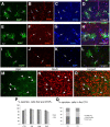
Figure 1.
Figure 1.
Reporter expression is initiated in cortical GFAP+ cells and not in neurons. GCE;R26R and GCE;CAG–EGFP hypoxic mice were analyzed at P15 after tamoxifen injections at P12–P14. A–H, EGFP/GFAP/S100β triple immunostaining; arrows point to corresponding double- and triple-stained cells. Arrowheads in E–H point to a reporter+/S100β+ astroglial cell that has undetectable GFAP expression. I–L, EGFP/Sox2 double immunostaining (DAPI in blue) showing that reporter+ cells express the pluripotency transcription factor Sox2 in the cerebral cortex. M–O, β-gal/NeuN double immunostaining shows that β-gal+ cells do not coexpress NeuN in the cerebral cortex. P shows that the percentage of reporter+ cells that are GFAP+ does not change between hypoxic- and normoxic-reared mice across brain regions. Q shows that all reporter+ cells in the cortex expressed GFAP, S100β, or both (n = 4 mice per condition). Images are 1 μm ApoTome single optical sections. CTX, Cortex; DG, dentate gyrus; WM, white matter; NX, normoxic; HX, hypoxic. Scale bar: A–O, P, 10 μm; Q, 50 μm.
Figure 2.
Figure 2.
Oligodendrocyte precursors cells arise from fate-mapped GFAP+ cells at P15. GCE mice carrying the EGFP reporter were analyzed at P15 after tamoxifen injections at P12–P14. A, Triple staining for EGFP, NG2, and PDGFRα in the P15 normoxic cerebral cortex. B, Stereological quantification showing that ∼10% of reporter+ cells express oligodendrocyte progenitor markers at P15 in normoxic mice; reporter+ cells express no mature oligodendrocyte markers at this stage. CTX, Cortex; WM, white matter. C, Cre/NG2 double immunostaining (DAPI in blue) in the cortex, demonstrating that Cre expression is not seen in NG2+ cells. D, Cre/GFAP double immunostaining (DAPI in blue) in the subcortical white matter and rostral migratory stream (RMS), demonstrating that Cre+ cells are GFAP+; percentages of Cre+ cells that express GFAP across different brain regions are shown. Ant CTX, Anterior cortex.
Figure 3.
Figure 3.
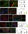
GFAP+ cells give rise to increased numbers of oligodendrocyte precursors cells and mature oligodendrocytes in hypoxic mice. GCE mice carrying the R26R or the EGFP reporter were analyzed at P35, 3 weeks after tamoxifen injections at P12–P14. A–C, β-gal/NG2 immunostaining showing a typical NG2+ cell expressing the Cre reporter in cortex. Quantification in D shows that, by P35, both NG2+ oligodendrocyte progenitors and mature Rip/CC1+ oligodendrocytes are reporter+. E–G, Example of β-gal/CC1+ cells in the white matter (WM). H, Density of β-gal/CC1+ cells in white matter (WM) of normoxic (Nx) and hypoxic (Hx) animals. I–K, Example of β-gal/Rip+ cell in cortex. L, Density of β-gal/Rip+ cells in cortex of normoxic and hypoxic animals. Images are 1 μm ApoTome single optical sections. Arrows point to reporter+ cells that are double positive. Scale bar, 20 μm.
Figure 4.
Figure 4.
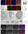
GFAP+ cells generate cortical neurons. GCE;R26R and GCE;CAG–GFP mice reared under normoxic (Nx) or hypoxic (Hx) conditions as indicated were analyzed at P35 (A–J) or P47 (K–Z) after tamoxifen injections at P12–P14. Sections were double immunostained with β-gal/NeuN (A–D), EGFP/NeuN (G–I), or single stained with β-gal followed by DAB detection (F). Some cortical neurons were NeuN+/reporter+ (arrows) and thus progeny of GFAP+ cells. The percentage of β-gal+ cells that express NeuN, NG2, APC, and GFAP in the cerebral cortex (CTX) is shown in E. The density of NeuN/β-gal+ cells is significantly increased in the cortex of hypoxic-reared animals (J). Images A–C are 1 μm ApoTome single optical sections, and D is a projection of six images spaced 1 μm apart. Scale bar, 20 μm. K–R, Newly generated neurons are synaptically connected in the cerebral cortex. GCE;CAG–EGFP mice reared under hypoxia were analyzed by EM after EGFP immunolabeling. Note EGFP immunoprecipitate in astrocyte end feet (N), dendrite (arrow in O), spines (arrows in P), and vesicle-containing axon terminals (double arrow in Q, R) apposed to synaptic thickening in spines. S–Z, EGFP/NeuN/Tbr1 triple immunostaining, demonstrating instances of reporter+/NeuN+ cells that are Tbr1+ (S–V) and reporter+ cells of neuronal morphology that are Tbr1 and NeuN (W–Z). Images are 1 μm ApoTome single optical sections.
Figure 5.
Figure 5.
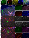
Hypoxia-induced amplification of neuronal progenitors derived from GFAP+ precursors in the SVZ and white matter. GCE;R26R (A–C) and GCE;CAG–EGFP (D–O) mice were tamoxifen injected at P12–P14 and analyzed at P35. A–C, In hypoxia-reared animals, the total number of Dcx+ neuronal progenitors and those Dcx+ cells that coexpress β-gal increased in the SVZ. D–H, Tbr2-immunostained cells were observed in the dorsal SVZ and RMS, glomerular layer (GL) of the OB, and boundary of cortex (CTX) with white matter (WM). The Tbr2+ cells in the white matter were Hu negative and appeared to undergo chain migration toward the cortex. I–L, Tbr2+/reporter+ cells (arrows), which arise from fate-mapped GFAP+ cells, are increased in the SVZ and WM of hypoxic mice. M–O, Tbr2+/BrdU+ cells (arrows) increased in the SVZ of hypoxic mice, although the proportion of Tbr2+ cells that incorporated BrdU (labeling index) was not significantly changed. Images are 1 μm ApoTome single optical sections. Hx, Hypoxic; Nx, normoxic.
Figure 6.
Figure 6.
Cortical GFAP+ cells are able to generate mature astrocytes and neurons. GCE; CAG–GFP mice were reared in hypoxia and then administered intracortical tamoxifen at P20. A–E, Four days after tamoxifen injection, Cre protein expression is observed in GFAP+, reporter+ cells adjacent to the injection tract in the cortex. F–J, Three weeks after tamoxifen administration, mature neurons (arrow) and astrocytes (arrowhead) were observed in the cortex of hypoxic animals, several millimeters distal from the point of injection. K–T, Mature neurons (K–O) and astrocytes (P–T) were still observed at 6 weeks after tamoxifen injection of hypoxic animals, suggesting long-term survival of these newly generated cells. Scale bars: (in A) AE, 10 μm; (in P) FJ, 40 μm, KT, 20 μm; GJ, LO, and QT are 50% reductions of F, K, and P, respectively.
Figure 7.
Figure 7.
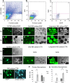
Cortical GFAP+ cells tagged in vivo with EGFP in GCE mice can generate neurospheres in hypoxic but not normoxic mice. A–C, Analysis of NG2+/GFAP+/GFP+ cells by flow cytometry. Dot plots of dissociated cells from GCE;CAG–EGFP transgenic mouse cortices in forward and side scatter with a polygon indicating the gate selecting the viable cells (A); GFP+ cells were gated based on the negative control cells (B). GFP+ cells were further analyzed according to fluorescence intensity for GFAP–PE (x-axis) and NG2–APC (y-axis) (C). D–I, Neurosphere (NS)-like cell clusters were obtained from the SVZ of both normoxic (Nx)- and hypoxic (Hx)-reared mice at P15. J–M, Neurosphere-like cell clusters were obtained from the hypoxic, but not the normoxic, cerebral cortex (CTX) at P15. N, O, Single green primary neurospheres from the hypoxic cortex dissociated and plated at clonal dilution generated exclusively green neurospheres, suggesting that a GFAP+ cell or its progeny were able to self-renew. P, Q, The total number of cell clusters (>80 μm in diameter) harvested from the different region was quantified 10–14 d after incubation (n = 12 per condition). For the SVZ, in vivo hypoxia increased the number of secondary neurospheres; for the cortex, hypoxia was required for both primary and secondary neurosphere formation. *p < 0.05; **p < 0.001.
Figure 8.
Figure 8.
Immunocytochemical analysis of differentiated EGFP+ neurospheres generated from hypoxic cortex. A–D and E–H, Magnified views of single neurospheres cultured for 10 d under differentiation conditions exhibiting MAP2 neurons (arrows) and GFAP+ astrocytes (arrowheads). I–L and M–P, Magnified views of single neurospheres exhibiting βIII-tubulin+ neuronal (arrows) and NG2+ OPCs (arrowheads). Q, Quantification of the percentages of EGFP+ neurospheres (ns) containing MAP2+ neurons, GFAP+ astrocytes, or both (n = 40 neurospheres analyzed). nd, Not determined. R, Quantification of the percentages of EGFP+ neurospheres containing βIII-tubulin+ neurons and NG2+ oligodendrocyte progenitors or both (n = 40 neurospheres analyzed). All neurospheres are from hypoxic cortex.
Figure 9.
Figure 9.
Exposure to low O2 in vitro maintains pluripotent fate potential in cortical GFAP+ cells. A, Scheme of the experimental paradigm. B, EGFP+ cells dissociated from secondary neurospheres exhibit astroglial phenotype and were directly apposed to Sox2-, NG2-, or MAP2-immunoreactive cells that were weakly EGFP+, suggesting a progenitor–progeny relationship. C, Primary neurospheres obtained under 5% O2 exhibit smaller size. D, Culture in 21% O2 for 3 d elicited a small increase in apoptotic cells as detected by caspase-3 immunostaining (D, arrow) compared with culturing in 5% O2. Hx, Hypoxic; Nx, normoxic. E, F, Tertiary neurospheres continuously grown in either 21 or 5% O2 were plated onto an adhesive substrate and grown under differentiating conditions. Triple MAP2/O1/GFAP immunostaining revealed that >75% of the neurospheres cultured in 5% O2 were able to generate neurons, astrocytes, and oligodendrocytes, whereas none of the neurospheres cultured in 21% O2 were neurogenic, and 80% could give rise to astrocytes only.

References

    1. Arvidsson A, Collin T, Kirik D, Kokaia Z, Lindvall O. Neuronal replacement from endogenous precursors in the adult brain after stroke. Nat Med. 2002;8:963–970. - PubMed
    1. Berninger B. Making neurons from mature glia: a far-fetched dream? Neuropharmacology. 2010;58:894–902. - PubMed
    1. Brenner M, Kisseberth WC, Su Y, Besnard F, Messing A. GFAP promoter directs astrocyte-specific expression in transgenic mice. J Neurosci. 1994;14:1030–1037. - PMC - PubMed
    1. Brill MS, Ninkovic J, Winpenny E, Hodge RD, Ozen I, Yang R, Lepier A, Gascón S, Erdelyi F, Szabo G, Parras C, Guillemot F, Frotscher M, Berninger B, Hevner RF, Raineteau O, Götz M. Adult generation of glutamatergic olfactory bulb interneurons. Nat Neurosci. 2009;12:1524–1533. - PMC - PubMed
    1. Buffo A, Rite I, Tripathi P, Lepier A, Colak D, Horn AP, Mori T, Götz M. Origin and progeny of reactive gliosis: a source of multipotent cells in the injured brain. Proc Natl Acad Sci U S A. 2008;105:3581–3586. - PMC - PubMed

Publication types

LinkOut - more resources