. 2011 Jun 22;31(25):9254-63.
doi: 10.1523/JNEUROSCI.1543-11.2011.

Prefrontal and monoaminergic contributions to stop-signal task performance in rats

Affiliations

Prefrontal and monoaminergic contributions to stop-signal task performance in rats

Andrea Bari et al. J Neurosci. .

Abstract

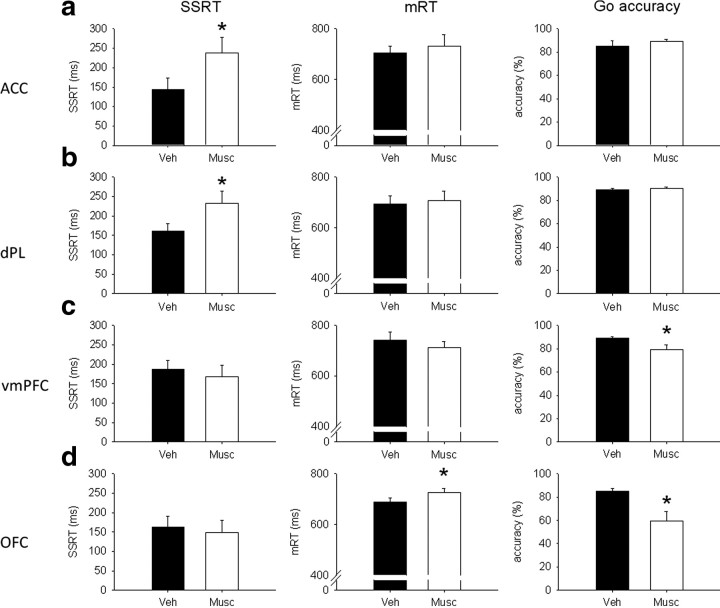
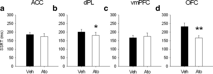
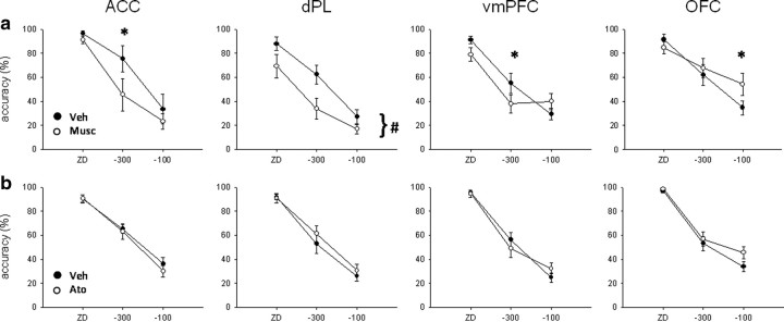
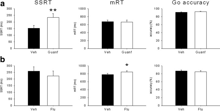
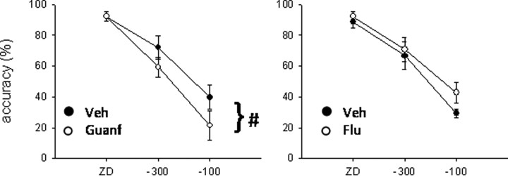
Defining the neural and neurochemical substrates of response inhibition is of crucial importance for the study and treatment of pathologies characterized by impulsivity such as attention-deficit/hyperactivity disorder and addiction. The stop-signal task (SST) is one of the most popular paradigms used to study the speed and efficacy of inhibitory processes in humans and other animals. Here we investigated the effect of temporarily inactivating different prefrontal subregions in the rat by means of muscimol microinfusions on SST performance. We found that dorsomedial prefrontal cortical areas are important for inhibiting an already initiated response. We also investigated the possible neural substrates of the selective noradrenaline reuptake inhibitor atomoxetine via its local microinfusion into different subregions of the rat prefrontal cortex. Our results show that both orbitofrontal and dorsal prelimbic cortices mediate the beneficial effects of atomoxetine on SST performance. To assess the neurochemical specificity of these effects, we infused the α2-adrenergic agonist guanfacine and the D(1)/D(2) antagonist α-flupenthixol in dorsal prelimbic cortex to interfere with noradrenergic and dopaminergic neurotransmission, respectively. Guanfacine, which modulates noradrenergic neurotransmission, selectively impaired stopping, whereas blocking dopaminergic receptors by α-flupenthixol infusion prolonged go reaction time only, confirming the important role of noradrenergic neurotransmission in response inhibition. These results show that, similar to humans, distinct networks play important roles during SST performance in the rat and that they are differentially modulated by noradrenergic and dopaminergic neurotransmission. This study advances our understanding of the neuroanatomical and neurochemical determinants of impulsivity, which are relevant for a range of psychiatric disorders.


Conflict of interest statement

Conflict of interest: T.W.R. discloses consultancy for Eli Lilly & Co., Pfizer, Cambridge Cognition, and research grants from Lundbeck, Pfizer, and Eli Lilly & Co.

Figures

Figure 1.
Figure 1.
a, Schematic illustration of the SST structure. A standard session consists of 210 trials to be completed within 30 min. On go trials (80% of total trials), the left and right levers have to be pressed in rapid sequence within the duration of the LH (1.2 s) to receive a reward, which is delivered in the central food well. On the remaining 20% of the trials (stop trials), an auditory stop signal will be played after the left lever has been pressed and after a variable SSD, which is calculated from each rat's mRT: ZD, mRT −300 and −100 ms. The presentation of the stop signal instructs the animal that the ongoing response to the right lever has to be inhibited to obtain the reward. Triangles represent the approximate time of presentation of the stop signal during the go response. b, Representative inhibition function obtained by plotting the SSDs against the probability of successful response inhibition. SSDs for the experimental phase (−300 and −100 ms) are chosen from the central part of the inhibition function, which is the most informative part both empirically and theoretically (Logan, 1994; Band et al., 2003).
Figure 2.
Figure 2.
Schematic representation of the position of the injector tips in experiment 1 (muscimol microinfusions; 0.5 μg/0.5 μl per side) as revealed by histological analysis. The sagittal view approximately shows the areas targeted by injectors on the sagittal plane. Open triangles, ACC (n = 4); filled circles, dPL (n = 12); open circles, vmPFC(n = 10); filled triangles, OFC (n = 9). Drawings adapted from Paxinos and Watson (1998).
Figure 3.
Figure 3.
Schematic representation of the position of the injector tips in experiment 2 as revealed by histological analysis. The sagittal view approximately shows the areas targeted by atomoxetine (0.1 μg/0.5 μl per side) microinjections on the sagittal plane. Open triangles, ACC (n = 14); filled circles, dPL (n = 13); open circles, vmPFC (n = 13); filled triangles, OFC (n = 11). Drawings adapted from Paxinos and Watson (1998).
Figure 4.
Figure 4.
Schematic representation of the position of the injector tips in experiment 3 as revealed by histological analysis. Guanfacine (a) (0.005 μg/0.5 μl per side) and α-flupenthixol (b) (15 μg/0.5 μl per side) were microinfused into the dPL (n = 7 for both). Drawings adapted from Paxinos and Watson (1998).
Figure 5.
Figure 5.
Effects of 0.5 μg/0.5 μl per side of muscimol (Musc) in ACC (a), dPL (b), vmPFC (c), and OFC (d) on SSRT (left), mRT (middle), and go accuracy (right). Muscimol infused into the ACC or dPL prolonged SSRT without affecting go-related measures. The same drug infused into the vmPFC or OFC impaired go-related measures disrupting global performance on the task. In these conditions of very low go accuracy (OFC), SSRT estimates may not be reliable. Veh, Vehicle. *p < 0.05.
Figure 6.
Figure 6.
Effects of 0.1 μg/0.5 μl per side of atomoxetine in ACC (a), dPL (b), vmPFC (c), and OFC (d) on SSRT. The selective norepinephrine reuptake inhibitor atomoxetine (Ato) in dPL and OFC improved response inhibition decreasing SSRT, without significant effects on go-related measures (data not shown). The same drug infused into the ACC or vmPFC did not have any effect on SSRT and other stop-signal task variables. Veh, Vehicle. *p < 0.05 and **p < 0.01.
Figure 7.
Figure 7.
Effect of muscimol (a; Musc) and atomoxetine (b; Ato) infusions into the ACC, dPL, vmPFC, and OFC. Muscimol (0.5 μg/0.5 μl per side) significantly impaired stop accuracy in ACC and dPL. In this latter region, stop accuracy was decreased across SSDs, whereas in the former, decomposition of the interaction revealed a significant effect only at −300 ms. Effects in OFC and vmPFC are likely to be caused by the effect of the drug on go accuracy (Fig. 5). Atomoxetine (0.1 μg/0.5 μl per side) did not affect significantly stop accuracy in any PFC subregion at the dose tested, suggesting that its effects are selective to the SSRT. Veh, Vehicle. *p < 0.05, simple main effect analysis; #p < 0.01, main effect only.
Figure 8.
Figure 8.
Effects of guanfacine (a; Guanf) and α-flupenthixol (b; Flu) microinfusions into the dPL on SSRT, mRT, and go accuracy. The α2A-adrenergic agonist guanfacine (0.005 μg/0.5 μl per side) selectively prolonged SSRT, whereas the mixed D1/D2 antagonist α-flupenthixol (15 μg/0.5 μl per side) prolonged go reaction time, without any effect on stopping. Veh, Vehicle. *p < 0.05 and **p < 0.01.
Figure 9.
Figure 9.
Effects of guanfacine (left; Guanf) and α-flupenthixol (right; Flu) on stop accuracy. Guanfacine infusion (0.005 μg/0.5 μl per side) significantly impaired stop accuracy, whereas α-flupenthixol (15 μg/0.5 μl per side) did not have any significant effect on this measure. Veh, Vehicle. #p < 0.05, main effect only.

References

    1. Aoki C, Venkatesan C, Go CG, Forman R, Kurose H. Cellular and subcellular sites for noradrenergic action in the monkey dorsolateral prefrontal cortex as revealed by the immunocytochemical localization of noradrenergic receptors and axons. Cereb Cortex. 1998;8:269–277. - PubMed
    1. Arnsten AF, Goldman-Rakic PS. Selective prefrontal cortical projections to the region of the locus coeruleus and raphe nuclei in the rhesus monkey. Brain Res. 1984;306:9–18. - PubMed
    1. Arnsten AF, Pliszka SR. Catecholamine influences on prefrontal cortical function: relevance to treatment of attention deficit/hyperactivity disorder and related disorders. Pharmacol Biochem Behav. 2011 doi: 10.1016/j.pbb.2011.01.020. - DOI - PMC - PubMed
    1. Aron AR, Robbins TW, Poldrack RA. Inhibition and the right inferior frontal cortex. Trends Cogn Sci. 2004;8:170–177. - PubMed
    1. Aron AR, Behrens TE, Smith S, Frank MJ, Poldrack RA. Triangulating a cognitive control network using diffusion-weighted magnetic resonance imaging (MRI) and functional MRI. J Neurosci. 2007;27:3743–3752. - PMC - PubMed

Publication types

Substances

LinkOut - more resources