. 2011 Jun 22;31(25):9279-88.
doi: 10.1523/JNEUROSCI.6606-10.2011.

Spontaneous age-related neurite branching in Caenorhabditis elegans

Affiliations

Spontaneous age-related neurite branching in Caenorhabditis elegans

Elizabeth M H Tank et al. J Neurosci. .

Abstract

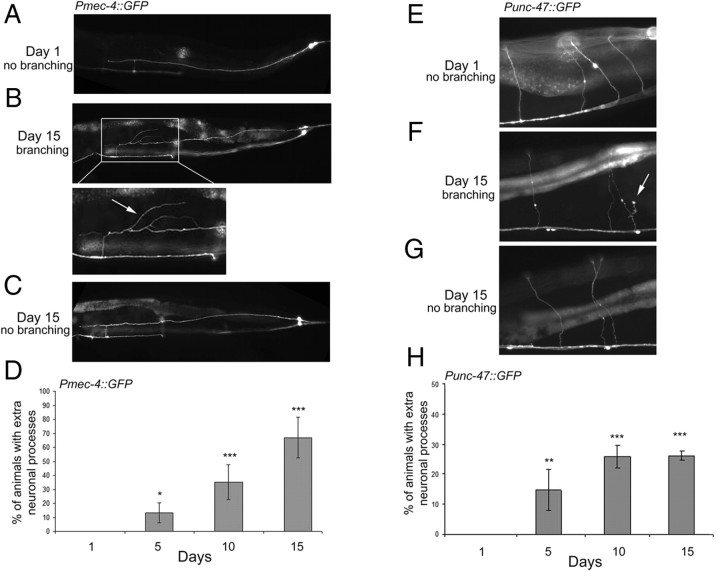
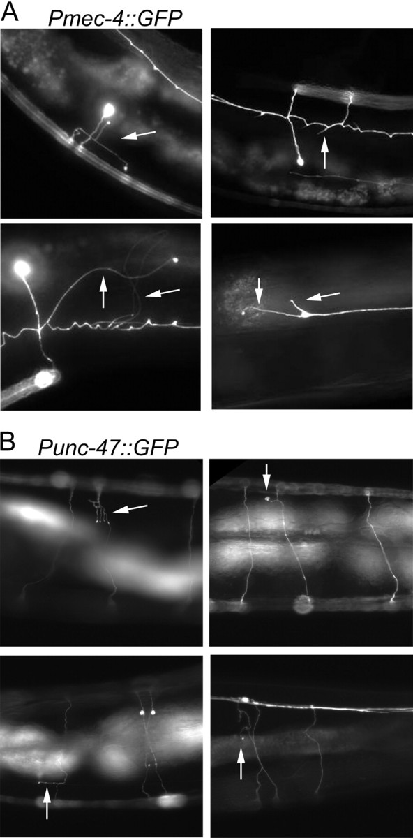
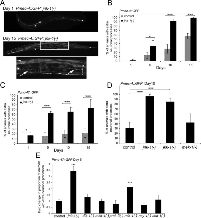
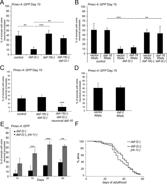
The analysis of morphological changes that occur in the nervous system during normal aging could provide insight into cognitive decline and neurodegenerative disease. Previous studies have suggested that the nervous system of Caenorhabditis elegans maintains its structural integrity with age despite the deterioration of surrounding tissues. Unexpectedly, we observed that neurons in aging animals frequently displayed ectopic branches and that the prevalence of these branches increased with time. Within age-matched populations, the branching of mechanosensory neurons correlated with decreased response to light touch and decreased mobility. The incidence of branching was influenced by two pathways that can affect the rate of aging, the Jun kinase pathway and the insulin/IGF-1 pathway. Loss of Jun kinase signaling, which slightly shortens lifespan, dramatically increased and accelerated the frequency of neurite branching. Conversely, inhibition of the daf-2 insulin/IGF-1-like signaling pathway, which extends lifespan, delayed and suppressed branching, and this delay required DAF-16/FOXO activity. Both JNK-1 and DAF-16 appeared to act within neurons in a cell-autonomous manner to influence branching, and, through their tissue-specific expression, it was possible to disconnect the rate at which branching occurred from the overall rate of aging of the animal. Old age has generally been associated with the decline and deterioration of different tissues, except in the case of tumor cell growth. To our knowledge, this is the first indication that aging can potentiate another form of growth, the growth of neurite branches, in normal animals.


Figures

Figure 1.
Figure 1.
Aged worms develop new neurite branches. A, A representative fluorescent image of a day 1 adult Pmec-4::GFP animal with structurally intact touch neurons. In all images, the tail of the animal is to the right. B, A representative fluorescent image of a day 15 adult Pmec-4::GFP animal with extra neurite branches. Bottom, Higher magnification of boxed area; the arrow highlights the ectopic neurite branches. C, A representative fluorescent image of a day 15 adult Pmec-4::GFP animal exhibiting morphologically intact neurons. D, Neurite branching increases during aging. Pmec-4::GFP animals were evaluated at each time point for the presence of extra neuronal processes in any one of the six touch neurons. The error bars represent the SD. n ≥ 3 experiments (χ2 test compared with day 1, *p < 0.01, ***p < 0.0001). E, A representative fluorescent image of GABAergic neurons in a day 1 adult Punc-47::GFP animal with normal commissures. F, A representative fluorescent image of a day 15 adult Punc-47::GFP animal with an extra neurite branch emanating from the commissure (arrow). G, A representative fluorescent image of a day 15 adult Punc-47::GFP animal exhibiting normal commissures (arrowheads). H, Neurite branches increase in GABAergic neurons during aging. Punc-47::GFP animals were evaluated at each time point for the presence of extra neuronal processes in commissures. n ≥ 2 experiments. (χ2 test compared with day 1, **p < 0.001, ***p < 0.0001).
Figure 2.
Figure 2.
Representative images of age-associated extra neurite branches. Pmec-4::GFP-expressing animals (A) and Punc-47::GFP expressing animals (B) displaying extra neurite branches (arrows).
Figure 3.
Figure 3.
Loss of jnk-1 enhances neurite branching with age. A, A representative fluorescent image of a day 1 adult Pmec-4::GFP; jnk-1(gk7) mutant animal (top). A representative fluorescent image of a day 15 adult Pmec-4::GFP; jnk-1(gk7) mutant animal exhibiting extra neurite branches (bottom). Bottom, Magnification of boxed area. Arrows indicate extra neurite branches. B, jnk-1 mutant worms display more neurite branches in touch neurons than do control animals. Control (Pmec-4::GFP) and Pmec-4::GFP; jnk-1(gk7) mutant animals were evaluated at each time point for the presence of extra neurite branches. The error bars represent the SD (χ2 test, *p < 0.01, ***p < 0.0001). C, jnk-1(−) mutant worms exhibit more neurite branches in GABAergic neurons than do control animals. Control (Punc-47::GFP) and Punc-47::GFP; jnk-1(gk7) mutant animals were evaluated at each time point for the presence of extra neurite branches. n ≥ 2 experiments. (χ2 test, *p < 0.01, ***p < 0.0001). D, Touch neurons in day 10 adult control (Pmec-4::GFP), jnk-1(gk7), jkk-1(km2), and mek-1(ks54) animals were analyzed for the presence of extra neurite branches. (χ2 test, ***p < 0.0001). E, GABAergic neurons in day 5 adult control (Punc-47::GFP), jnk-1(gk7), dlk-1(ju476), mkk-4(ju91), pmk-3(ok169), mlk-1(ok2471), nsy-1(ok593), and sek-1(km4) were analyzed for the presence of extra neurite branches. Because of the use of two different Punc-47::GFP alleles, the data were normalized to control (ANOVA, ***p < 0.0001 compared with control). This analysis was performed at day 5 of adulthood because the jnk-1(−) mutant already exhibited a dramatic increase in branching compared with control.
Figure 4.
Figure 4.
Insulin/IGF-1 signaling promotes extra neurite branching. A, The extra neurite branches were suppressed by loss of daf-2 in a daf-16-dependent manner. The touch neurons were analyzed in day 10 adult control (Pmec-4::GFP), daf-2(e1370) mutants, daf-16(mu86) mutants, and daf-16(mu86); daf-2(e1370) double mutants (χ2 test, **p < 0.001, ***p < 0.0001). B, daf-16 must be depleted in neurons to relieve daf-2-dependent suppression of branching. Touch neurons of daf-2(e1370) animals were analyzed at day 10 after growth on control (vector-only) RNAi bacteria and bacteria expressing dsRNA against daf-16. As predicted, daf-16; daf-2 double mutants fed daf-16--RNAi bacteria exhibited wild-type levels of neurite branching (χ2 test, **p < 0.001, ***p < 0.0001). C, Neuronal expression of daf-16 suppresses branching in daf-16(−); daf-2(−) mutant animals. The touch neurons were observed on day 10 of adulthood in control (Pmec-4::GFP), daf-16(mu86); daf-2(e1370), and daf-16(mu68); daf-2(e1370); Punc-119::GFP::daf-16 animals [χ2 test, ***p < 0.001 compared with control or daf-16(−); daf-2(−)]. D, daf-2 must be depleted from neurons to suppress branching. Pmec-4::GFP animals were analyzed on day 15 of adulthood after growth on control (vector-only) RNAi bacteria or bacteria expressing dsRNA against daf-2. E, daf-2 and jnk-1 mutations may act in different pathways to influence neurite branches. The touch neurons of daf-2(e1370) and daf-2(e1370); jnk-1(gk7) were evaluated on days 10, 15, 20, and 40 for the presence of neurite branches. Loss of jnk-1 enhanced the percentage of animals displaying branches in daf-2 mutant animals but not to the same extent as loss of jnk-1 in otherwise wild-type animals. n ≥ 2 experiments (χ2 test, **p < 0.001, ***p < 0.0001). F, Loss of jnk-1 did not shorten the daf-2 mutant lifespan. Lifespan analysis was performed on Pmec-4::GFP; daf-2(e1370) and Pmec-4::GFP; daf-2(e1370); jnk-1(gk7) animals. Mean lifespan for daf-2(−): 35.2 d. Mean lifespan for daf-2(−); jnk-1(−): 37.6 d. These lifespans were not significantly different.
Figure 5.
Figure 5.
Not all longevity mutations suppress neurite branching. A, Long-lived eat-2 mutants have similar wild-type levels of neurite branching, yet long-lived clk-1 mutants have suppressed branching. The touch neurons of day 15 control (Pmec-4::GFP), clk-1(qm30), and eat-2(ad1116) mutant animals were evaluated for the presence of extra neurite branches (χ2 test compared with control, ***p < 0.0001). B, A lifespan curve of control (Pmec-4::GFP), Pmec-4::GFP; clk-1(qm30), and Pmec-4::GFP; eat-2(ad1116) mutant animals, performed in parallel with the branching analysis in A. The mean lifespans were as follows: control at 18.5 d, clk-1(−) at 25.4 d, and eat-2(−) at 27.4 d (Mantel–Cox log-rank test, p < 0.0001).

References

    1. Adachi H, Fujiwara Y, Ishii N. Effects of oxygen on protein carbonyl and aging in Caenorhabditis elegans mutants with long (age-1) and short (mev-1) life spans. J Gerontol A Biol Sci Med Sci. 1998;53:B240–B244. - PubMed
    1. Björkblom B, Ostman N, Hongisto V, Komarovski V, Filén JJ, Nyman TA, Kallunki T, Courtney MJ, Coffey ET. Constitutively active cytoplasmic c-Jun N-terminal kinase 1 is a dominant regulator of dendritic architecture: role of microtubule-associated protein 2 as an effector. J Neurosci. 2005;25:6350–6361. - PMC - PubMed
    1. Bogoyevitch MA, Kobe B. Uses for JNK: the many and varied substrates of the c-Jun N-terminal kinases. Microbiol Mol Biol Rev. 2006;70:1061–1095. - PMC - PubMed
    1. Braeckman BP, Houthoofd K, De Vreese A, Vanfleteren JR. Apparent uncoupling of energy production and consumption in long-lived Clk mutants of Caenorhabditis elegans. Curr Biol. 1999;9:493–496. - PubMed
    1. Brugg B, Matus A. Phosphorylation determines the binding of microtubule-associated protein 2 (MAP2) to microtubules in living cells. J Cell Biol. 1991;114:735–743. - PMC - PubMed

Publication types