

Nodular regenerative hyperplasia of the liver: coral atoll-like lesions on ultrasound are characteristic in predisposed patients
- PMID: 21697407
- PMCID: PMC3473481
- DOI: 10.1259/bjr/17975057
Nodular regenerative hyperplasia of the liver: coral atoll-like lesions on ultrasound are characteristic in predisposed patients
Abstract
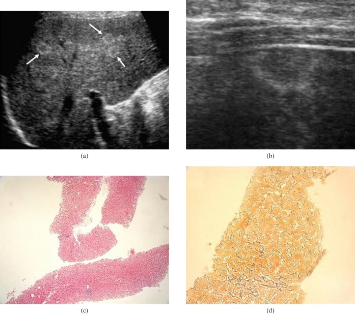
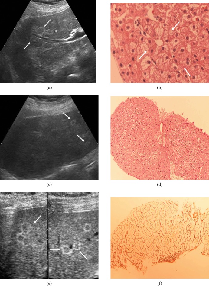
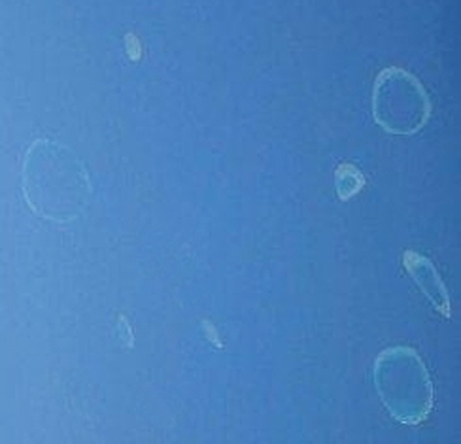
Nodular regenerative hyperplasia (NRH) is an uncommon liver disease characterised histologically by numerous small hyperplastic nodules that are not separated by fibrotic tissue. It is thought to be the result of obliterative vasculopathy, and it has been associated with chronic use of medications, toxic substances and a wide variety of systemic diseases. Imaging diagnosis of early-stage NRH remains problematic. The nodules are rarely discerned and their appearance and behaviour before and after contrast medium administration are heterogeneous and not specific. A review of the literature shows that ultrasound has succeeded on occasion in revealing small focal liver lesions in patients with NRH. To our knowledge, there has been no published data on the performance in this setting of last-generation ultrasound scanners and techniques such as contrast-enhanced ultrasound (CEUS). The question is an important one because abdominal ultrasound is widely used as a first-line imaging technique for the evaluation of liver disease, and this makes it particularly suitable as a potential tool for the early diagnosis of NRH. Owing to the prolonged subclinical period and the limited help provided by imaging, the diagnosis in vivo of NRH is currently frequently missed, and it is still made exclusively on the basis of liver biopsy. In conclusion, this report describes 4 cases of biopsy-proven NRH that have been diagnosed over the past 2 years by our group. All were characterised by known comorbidities that confer a predisposition to NRH and by a peculiar parenchymal ultrasound pattern that we refer to as the "atoll sign".
Figures
References
-
- Wanless IR. Micronodular transformation (nodular regenerative hyperplasia) of the liver: a report of 64 cases among 2,500 autopsies and a new classification of benign hepatocellular nodules. Hepatology 1990;11:787–97 - PubMed
-
- Dachman AH, Ros PR, Goodman ZD, Olmsted WW, Ishak KG. Nodular regenerative hyperplasia of the liver: clinical and radiological observations. AJR 1987;148:717–22 - PubMed
-
- Siegelman ES, Outwater EK, Furth EE, Rubin R. MR imaging of hepatic nodular regenerative hyperplasia. J Magn Reson Imaging 1995;5:730–2 - PubMed
-
- Ames JT, Federle MP, Chopra K. Distinguishing clinical and imaging features of nodular regenerative hyperplasia and large regenerative nodules of the liver. Clin Radiol 2009;64:1190–5 - PubMed
-
- Flejou JF, Barge J, Menu Y, Degott C, Bismuth H, Potet F, et al. Liver adenomatosis. An entity distinct form liver adenoma? Gastroenterology 1985;89:1132–8 - PubMed
Publication types
MeSH terms
Substances
LinkOut - more resources
Full Text Sources
Medical
Miscellaneous
