

Isolated intracranial Rosai-Dorfman disease mimicking meningioma in a child: a case report and review of the literature
- PMID: 21697409
- PMCID: PMC3473498
- DOI: 10.1259/bjr/15772106
Isolated intracranial Rosai-Dorfman disease mimicking meningioma in a child: a case report and review of the literature
Abstract
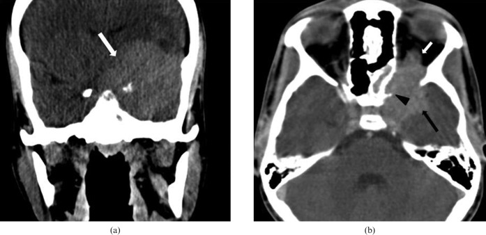
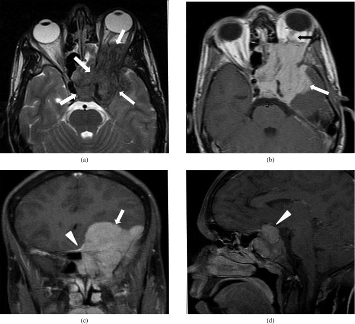
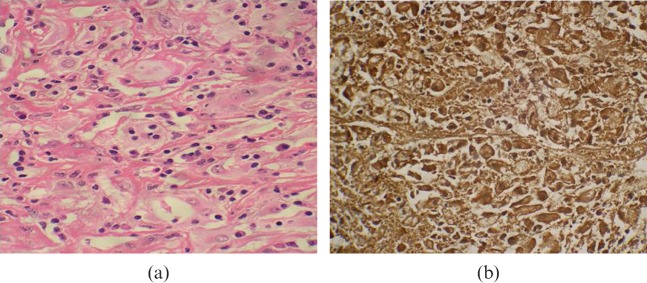
We report the first case of extensive involvement of isolated intracranial Rosai-Dorfman's disease (RDD) in a child. Our case is unique because it presents with involvement of the middle cranial fossa, cavernous sinus, pituitary gland, orbit, ethmoid and sphenoid sinuses. Previous cases of intracranial RDD in children have reported separate involvement of cavernous sinus, suprasellar region, and frontal and petroclival regions. Involvement of the pituitary gland has so far not been reported. A 14-year-old male presented with a medical history of loss of vision, raised erythrocyte sedimentation rate (ESR), and abnormal prolactin and cortisol levels. Radiologically the diagnosis was meningioma. The histopathological diagnosis was RDD with emperipolesis and S-100 positivity. RDD is a histiocytic proliferation of unknown aetiology, which commonly affects lymph nodes. Uncommonly it involves the extranodal sites and rarely the central nervous system (CNS). 80 cases of RDD involving CNS have been reported in the literature, and only 5 were in children. Although the definitive diagnosis of RDD disease is based on the histopathology report, it should be included in the differentials of a lesion mimicking meningioma especially in children.
Figures
References
-
- Rosai J, Dorfman RF. Sinus histiocytosis with massive lymphadenopathy: a newly recognized benign clinicopathological entity. Arch Pathol 1969;87:63–70 - PubMed
-
- Wan S, Teng X, Zhan R, Yu J, Gu J, Zhang K. Isolated intracranial Rosai-Dorfman disease mimicking suprasellar meningioma: case repot with review of the literature. J Int Med Re 2008;36:1134–9 - PubMed
-
- Theeler BJ, Keylock JB, Yoest SM. Teaching neuroimage: isolated intracranial Rosai-Dorfman disease mimicking a meningioma. Neurology 2008;70:e42. - PubMed
-
- Shaver EG, Robsamem SL, Yachnis AT, Sutton LN. Isolated extranodal sinus histiocytosis in a 5 year old boy: a case report. J Neurosurg 1993;79:769–73 - PubMed
-
- Woodcock R, Mandell J, Lipper M. Sinus histiocytosis (Rosai–Dorfman disease) of the suprasellar region: MR imaging findings—a case report. Radiology 1999;213:808–10 - PubMed
Publication types
MeSH terms
LinkOut - more resources
Full Text Sources
Miscellaneous
