. 2011 Aug 1;187(3):1150-6.
doi: 10.4049/jimmunol.1004077. Epub 2011 Jun 22.

Obstructive jaundice expands intrahepatic regulatory T cells, which impair liver T lymphocyte function but modulate liver cholestasis and fibrosis

Affiliations

Obstructive jaundice expands intrahepatic regulatory T cells, which impair liver T lymphocyte function but modulate liver cholestasis and fibrosis

Steven C Katz et al. J Immunol. .

Abstract

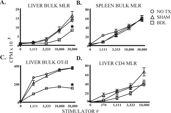
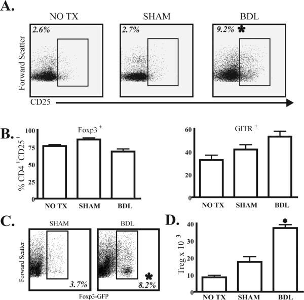
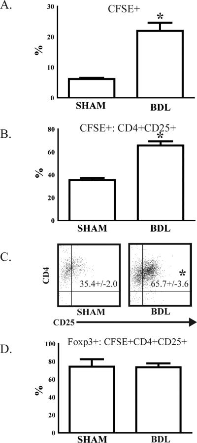
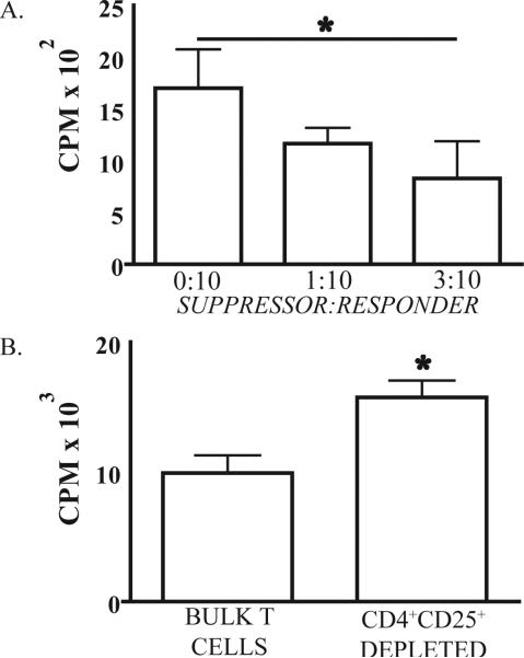
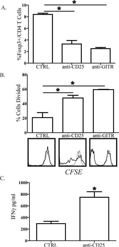
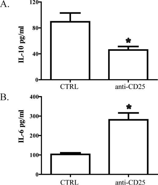
Although obstructive jaundice has been associated with a predisposition toward infections, the effects of bile duct ligation (BDL) on bulk intrahepatic T cells have not been clearly defined. The aim of this study was to determine the consequences of BDL on liver T cell phenotype and function. After BDL in mice, we found that bulk liver T cells were less responsive to allogeneic or syngeneic Ag-loaded dendritic cells. Spleen T cell function was not affected, and the viability of liver T cells was preserved. BDL expanded the number of CD4(+)CD25(+)Foxp3(+) regulatory T cells (Treg), which were anergic to direct CD3 stimulation and mediated T cell suppression in vitro. Adoptively transferred CD4(+)CD25(-) T cells were converted into Treg within the liver after BDL. In vivo depletion of Treg after BDL restored bulk liver T cell function but exacerbated the degrees of inflammatory cytokine production, cholestasis, and hepatic fibrosis. Thus, BDL expands liver Treg, which reduce the function of bulk intrahepatic T cells yet limit liver injury.


Figures

Figure 1
Figure 1. Liver bulk T cells are suppressed following BDL
B6 mice underwent bile duct ligation (BDL), sham laparotomy (SHAM), or no treatment (NO TX). After 7 days, T cells were stimulated with DC. Bulk liver (A) but not spleen (B) T cell function in MLR was suppressed by BDL. Similar findings were noted when liver T cells from OT-II mice 7 days after BDL were stimulated with OVA-loaded syngeneic DC (C). The conventional CD4 (CD25-NK1.1-CD1d/αGalCer-γδ-) T cell response to allogeneic DC was not affected by BDL (D). Proliferation of T cells or DC cultured alone was negligible. Means and standard deviations are shown based on triplicate wells and the data are representative of 3 or more repetitions with 3 or more livers pooled per group. *p<0.05
Figure 2
Figure 2. BDL expands liver regulatory T cells in B6 mice
(A) Flow cytometry was used to determine the frequency of CD25+ lymphocytes among the liver CD4 T cell (CD3+CD4+NK-γδ-) population at 7 days following BDL. CD25 gating was based on negative isotype control staining. (B) To determine the proportion of the CD4+CD25+ liver T cell population that were actually Treg, we measured the levels of FoxP3 and GITR expression among these cells in all three groups. (C) Liver Treg expansion following BDL was confirmed in FoxP3-GFP mice, in which all FoxP3+ cells co-express GFP. We gated upon viable CD3+CD4+ liver T cells to determine the proportion of cells expressing GFP and hence FoxP3. (D) The total number of CD4+CD25+FoxP3+ cells per liver is shown. Percentages and absolute numbers are means from 3 animals per group and are representative of 2-3 repetitions. *p<0.05
Figure 3
Figure 3. Liver Treg expansion is due in part to conversion of CD4+ CD25- T cells
To determine if liver Treg were derived from CD4 T cell precursors following BDL, CD4+CD25-splenic T cells were isolated from B6 mice, labeled with CFSE, and then (1 × 10⁶) adoptively transferred via the portal vein at the time of BDL or SHAM. After 7 days, liver T cells were harvested and analyzed by flow cytometry. (A) The proportion of CFSE+ adoptively transferred cells among bulk (Thy1.2+) liver T cells was significantly higher following BDL. (B & C) To determine the proportion of adoptively transferred cells that adopted a Treg phenotype, we measured CD25 expression among CFSE+ cells. (D) FoxP3 expression was confirmed in isolated, converted CD4+CD25+ T cells. Representative of two repetitions with 3 mice per group. *p<0.05
Figure 4
Figure 4. Liver CD4+ CD25+ T cells from BDL treated mice have suppressive properties
(A) 1 × 10⁴ CD4+CD25- liver T cells and varying numbers of CD4+CD25+ T cells from BDL mice were cultured with 5 × 10³ splenic DC from Balb/c mice in a 3 day mixed leukocyte reaction (MLR). CD4+CD25- and CD4+CD25+ liver T cells were purified from the same jaundiced animals. (B) From BDL mice, 5 × 10⁴ bulk liver T cells or bulk liver T cells sorted to exclude CD4+CD25+ cells were cultured with 3 × 10⁴ splenic DC from Balb/c mice in a 3 day MLR. Proliferation of T cells or DC cultured alone was negligible. Mean and standard deviation are shown based on triplicate wells and the data are representative of 2 repetitions with three or more livers pooled in each group. *p<0.05
Figure 5
Figure 5. In vivo depletion of regulatory T cells restores bulk liver T cell function following BDL
B6 mice were treated with 100μg of anti-CD25 (PC61) or anti-GITR (DTA1) i.p. on days -1, 0, 5, and 7 in relation to BDL. On D8, livers were harvested and Thy1.2+ bulk T cells were isolated for confirmation of depletion or labeled with CFSE and co-cultured with allogeneic splenic DC. After 72-96 hours of co-culture, T cells were analyzed by flow cytometry to measure the (A) percentage of FoxP3+ cells among CD4 T cells remaining after depletion and (B) proliferation by CFSE dissolution. For proliferation data, the bar graphs show percentage of cells that divided. On the histograms, solid lines represent T cells stimulated with DC and the dashed lines unstimulated T cells. (C) IFNγ in supernatants from the anti-CD25 depletion experiment was analyzed by cytometric bead array confirm enhanced T cell function following Treg depletion. Data are representative of 3 independent experiments.
Figure 6
Figure 6. Liver Treg depletion results in diminished IL-10 but increased IL-6 production from liver T cells following bile duct ligation
B6 mice were treated with 100μg of anti-CD25 (PC61) i.p. on days -1, 0, 5, and 7 in relation to BDL. On D8, liver bulk T cells were harvested and co-cultured with allogeneic splenic DC for 3-5 days. Supernatant was harvested and (A) IL-10 or (B) IL-6 were measured by cytometric bead array. Data are representative of two independent experiments.
Figure 7
Figure 7. Liver Treg depletion exacerbates biliary tract injury and cholestasis during obstructive jaundice
B6 mice were treated with 100μg of anti-CD25 (PC61) i.p. on days -1, 0, 5, and 7 in relation to BDL. On day 8, animals were sacrificed for blood collection and histologic assessment of liver tissue. When Treg were depleted, BDL resulted in significantly higher serum bilirubin (A) and alkaline phosphatase (B) levels. (C) Liver tissue analyzed with Mallory's trichrome stain to demonstrate collagen confirmed that Treg depletion resulted in excessive peri-portal fibrosis and infiltration with inflammatory cells. Images shown are at 100X (top row) and 200X magnification (bottom row).

References

    1. Bleier JI, Katz SC, Chaudhry UI, Pillarisetty VG, Kingham TP, 3rd, Shah AB, Raab JR, DeMatteo RP. Biliary obstruction selectively expands and activates liver myeloid dendritic cells. J Immunol. 2006;176:7189–7195. - PubMed
    1. Armstrong CP, Dixon JM, Duffy SW, Elton RA, Davies GC. Wound healing in obstructive jaundice. Br J Surg. 1984;71:267–270. - PubMed
    1. Bailey ME. Endotoxin, bile salts and renal function in obstructive jaundice. Br J Surg. 1976;63:774–778. - PubMed
    1. Jiang WG, Puntis MC. Immune dysfunction in patients with obstructive jaundice, mediators and implications for treatments. HPB Surg. 1997;10:129–142. - PMC - PubMed
    1. Greig JD, Krukowski ZH, Matheson NA. Surgical morbidity and mortality in one hundred and twenty-nine patients with obstructive jaundice. Br J Surg. 1988;75:216–219. - PubMed

Publication types

MeSH terms

Substances