. 2011 Sep;85(17):8870-83.
doi: 10.1128/JVI.00059-11. Epub 2011 Jun 22.

Hepatitis C virus stimulates the phosphatidylinositol 4-kinase III alpha-dependent phosphatidylinositol 4-phosphate production that is essential for its replication

Affiliations

Hepatitis C virus stimulates the phosphatidylinositol 4-kinase III alpha-dependent phosphatidylinositol 4-phosphate production that is essential for its replication

Kristi L Berger et al. J Virol. 2011 Sep.

Abstract

Phosphatidylinositol 4-kinase III alpha (PI4KA) is an essential cofactor of hepatitis C virus (HCV) replication. We initiated this study to determine whether HCV directly engages PI4KA to establish its replication. PI4KA kinase activity was found to be absolutely required for HCV replication using a small interfering RNA transcomplementation assay. Moreover, HCV infection or subgenomic HCV replicons produced a dramatic increase in phosphatidylinositol 4-phosphate (PI4P) accumulation throughout the cytoplasm, which partially colocalized with the endoplasmic reticulum. In contrast, the majority of PI4P accumulated at the Golgi bodies in uninfected cells. The increase in PI4P was not observed after infection with UV-inactivated HCV and did not reflect changes in PI4KA protein or RNA abundance. In an analysis of U2OS cell lines with inducible expression of the HCV polyprotein or individual viral proteins, viral polyprotein expression resulted in enhanced cytoplasmic PI4P production. Increased PI4P accumulation following HCV protein expression was precluded by silencing the expression of PI4KA, but not the related PI4KB. Silencing PI4KA also resulted in aberrant agglomeration of viral replicase proteins, including NS5A, NS5B, and NS3. NS5A alone, but not other viral proteins, stimulated PI4P production in vivo and enhanced PI4KA kinase activity in vitro. Lastly, PI4KA coimmunoprecipitated with NS5A from infected Huh-7.5 cells and from dually transfected 293T cells. In sum, these results suggest that HCV NS5A modulation of PI4KA-dependent PI4P production influences replication complex formation.


Figures
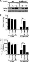
Fig. 1.
Fig. 1.
PI4KA kinase activity is required for HCV replication. Transcomplementation assays of Huh-7.5 cells were performed 86 h after siRNA electroporation, 72 h after pTrip-pseudoparticle infection with wild-type (WT) or kinase-deficient (KD) PI4KA, and 48 h after HCV infection. siRNAs included si-irrelevant (IRR) and si-PI4KA-endogenous (PI4KA-endg). (A) PI4KA protein expression was verified by immunoblot compared to a β-actin control. (B) HCV replication as measured by qRT-PCR and calculated relative to si-IRR-treated, vector-transduced cells. (C) Infectious virus production as measured by tissue culture infectious dose 50 (TCID50)/ml from limiting dilution analysis. For panels B and C, the data are expressed based on triplicates + the SEM in a log scale. **, P < 0.001.
Fig. 2.
Fig. 2.
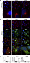
HCV infection increases PI4P production. (A) Confocal microscopy of PI4P and NS5A in Huh-7.5 cells at 48 h after treatment with infectious HCV (multiplicity of infection [MOI] = 0.2), UV-inactivated HCV, or electroporation with subgenomic (sg) HCV replicons (JFH-1 isolate). sgJFH-1 electroporated cells and null-electroporated cells were mixed 1:1 and seeded on coverslips for side-by-side comparison. PI4P (red) was detected by indirect immunofluorescence. NS5A (green) was directly detected using anti-NS5A-488-conjugated antibody staining. Nuclei were stained with DAPI. Scale bar, 20 μm. (B) Quantitation of PI4P fluorescence per cell from multiple images from triplicate coverslips. Averages + the SEM are shown. **, P < 0.001. (C) Detection of PI4P from lipid extracts of HCV- or mock-infected Huh-7.5 cells 48 h postinfection using PI4P mass strips, with findings representative of two experiments. Column a consists of three sets of extractions (1 to 3) from experimental samples. Column b consists of prespotted PI4P standards as follows (from top to bottom): 10, 5, 4, 2, 1, and 0.5 pmol.
Fig. 3.
Fig. 3.
PI4KA abundance is unaltered by HCV infection. Huh-7.5 cells were infected with UV-inactivated (UV-HCV) or infectious HCV (MOI = 3) and analyzed at indicated hours postinfection (HPI). (A) Total PI4KA mRNA was quantified by real-time RT-PCR from triplicate infected samples for each time point and calculated relative to a 2-h sample. The SEM is shown. ns, not significant. Results are representative of three experiments. (B) HCV RNA transcripts from the same samples as in panel A, similarly quantified. (C) Proteins analyzed at the indicated time points (in hours) postinfection by immunoblotting for PI4KA, NS5A, and β-actin.
Fig. 4.
Fig. 4.
Redistribution of PI4P by HCV infection. PI4P (red), GM130 (green), and calnexin (green) were detected by indirect immunofluorescence in Huh-7.5 cells either mock or HCV infected for 48 h (MOI = 3). Viral infection was detected by direct immunostaining for NS5A using anti-NS5A-488-conjugated antibody. PI4P fluorescence is normalized between images of mock-infected and infected cells for comparison (red = 166-ms exposure). Nuclei were stained with DAPI. *, Cell magnified in the bottom panel. Scale bar, 10 μm.
Fig. 5.
Fig. 5.
HCV NS5A enhances PI4P accumulation in stable U2OS cell lines with inducible HCV protein expression. U2OS osteosarcoma UHCV (polyprotein), UNS5A, and UNS5B cell lines were grown in either uninduced conditions (A) or induced expression conditions (B) and imaged 48 h postinduction. In both panels A and B, PI4P (red) and NS5B (green) were detected using indirect immunofluorescence. NS5A was directly detected using anti-NS5A-488-conjugated antibody. PI4P fluorescence imaging parameters were kept constant for all images in A and B (red = 100-ms exposure). *, Cell magnified in the bottom image in panels A and B. Arrowheads denote the foci of PI4P colocalized with NS5A. Arrows denote PI4P foci without NS5A. Nuclei were stained with DAPI. Scale bar, 20 μm. (C) Quantitation of images represented in panels A and B as the percent cells with PI4P foci redistributed throughout the cytosol from uninduced or induced cells, as verified by viral protein immunostaining. n, the total number of cells. The data were from two independent experiments for UHCV and UNS5A and one for UNS5B.
Fig. 6.
Fig. 6.
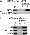
PI4KA coimmunoprecipitates with NS5A. (A) Huh-7.5 cells were electroporated with full-length HCV RNAs, and lysate was collected 48 h later from a 150-mm dish. Lysates were divided equally between immunoprecipitation reactions with anti-mouse IgG magnetic beads containing NS5A primary antibody (+Ab) or without (−Ab). NS5A and endogenous PI4KA (endg.PI4KA) were detected by immunoblotting (IB). The input represents 1% extract, and IP is 25% of the total pulldown; the results are representative of three independent experiments. (B) HEK 293T cells were cotransfected with HA-tagged PI4KA and untagged NS5A expression constructs, and lysates were collected at 48 h posttransfection, immunoprecipitated, and immunoblotted as in panel A. The PI4KA immunodetection in panel B represents both HA-tagged and endogenous proteins.
Fig. 7.
Fig. 7.
NS5A stimulates PI4KA activity. (A) HEK 293T cells were transfected with empty vector or plasmids encoding HA-tagged wild-type (WT) or a kinase-deficient (KD) mutant (K1792L) of PI4KA. PI4KA was immunoprecipitated (IP) using anti-HA antibody [IP (+)Ab; lanes 1 to 3] and verified by PI4KA immunoblotting (IB) compared to lysates incubated with beads alone without HA antibody [IP (−)Ab; lanes 4 to 5]. IP is 8% of total pulldown. (B) Immunoprecipitates of beads only or HEK 293T cells transfected with vector, HA-PI4KA-WT, or HA-PI4KA-KD were added to an in vitro kinase reaction in triplicate (see Materials and Methods). The concentrations of drugs used were as follows: 500 nM wortmannin (WORT) or 5 μM phenylarsine oxide (PAO) diluted in DMSO. The SEM is shown for all data points but is indiscernible for some samples. (C) Silver-stained 4 to 20% SDS-PAGE gel showing the purity of recombinant HisV5-NS5A and NS5BΔ21-His proteins. HA-PI4KA (∼230 kDa) was specifically immunoprecipitated by HA antibody, (+)Ab. The negative control, (−)Ab, consisted of immunoprecipitates from HA-PI4KA-transfected lysate lacking HA antibody. (D) The kinase assay activity of ∼25 ng of immunoprecipitated HA-PI4KA, or equivalent (−)Ab control, was measured after the addition of purified NS5A or NS5B (see Materials and Methods). 1×, 5×, and 25× represent the molar excesses of viral proteins compared to HA-PI4KA. For PI4KA inhibition, 5 μM PAO diluted in DMSO was used. Counts per minute (CPM) + the SEM represent results for triplicate reactions. *, P < 0.005 compared to empty buffer (none) from HA-PI4KA reactions. The results are representative of four experiments.
Fig. 8.
Fig. 8.
RNA interference analysis of PI4P production in UHCV cells. The U2OS cell line with inducible HCV polyprotein expression (UHCV) was treated with siRNAs targeting PI4KA, PI4KB, or irrelevant (IRR), followed by growth in either uninduced or induced media conditions. The data were collected 54 h after siRNA and at 32 h postinduction. (A) Immunoblot of PI4KA, PI4KB, and β-actin control of UHCV cells treated with siRNAs and grown under induction conditions. (B) PI4P (red) and PI4KB (green) were detected by indirect immunofluorescence for siRNAs and growth conditions indicated. For red and green channels, imaging parameters were constant between all images for comparison (exposure: red = 42 ms; green = 295 ms). A DAPI nuclear stain was performed. Scale bar, 20 μm.
Fig. 9.
Fig. 9.
PI4P induction by HCV polyprotein expression is dependent on PI4KA. U2OS UHCV cells were examined under the same conditions as in Fig. 8. (A and B) Indirect immunofluorescence of PI4P (red) in uninduced cells or cells induced for HCV polyprotein expression treated with irrelevant (IRR), PI4KA, or PI4KB siRNAs with direct detection of NS5A (green) using anti-NS5A-488 antibody (A) or with indirect detection of Golgi-marker GM130 (B). The labels for panel B are as described for panel A. Representative images are shown. Scale bar, 10 μm. Red and green fluorescence imaging parameters were kept constant among all images within each panel (exposure, panel A: red = 102 ms, green = 73 ms; exposure, panel B: red = 85 ms, green = 2,208 ms).
Fig. 10.
Fig. 10.
ER/cytosolic PI4P accumulation following HCV polyprotein expression is dependent on PI4KA. Quantitation is based on the experimental setup and microscopy shown in Fig. 8. Comparing uninduced versus induced growth conditions for each siRNA treatment, the number of UHCV cells with broad cytosolic PI4P distribution beyond GM130 (PI4P-ER/cytosolic) was quantified from multiple images represented in panel B. The number of NS5A-expressing cells in each siRNA-treated population was quantified from multiple images represented in panel A. The total number of cells counted (n) is indicated.
Fig. 11.
Fig. 11.
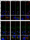
PI4KA is required for proper localization of HCV replicase proteins. The U2OS UHCV cell line was examined at 72 h after siRNA treatment, either si-irrelevant (si-IRR) or si-PI4KA, and at 48 h postinduction of HCV polyprotein expression. For (red) indirect immunofluorescence detection of PI4P, the cells were permeabilized with digitonin, whereas for the detection of NS5B and NS3, Triton X-100 and saponin were used. NS5A was directly detected using anti-NS5A-488-conjugated antibody (green). NS5A, NS5B, and NS3 were not detected under uninduced conditions (data not shown). For red channels, imaging parameters were constant between si-IRR and si-PI4KA within each antibody sample set. DAPI nuclear stain was included. Scale bar, 10 μm.

References

    1. Ahn J., et al. 2004. Systematic identification of hepatocellular proteins interacting with NS5A of the hepatitis C virus. J. Biochem. Mol. Biol. 37:741–748 - PubMed
    1. Amako Y., Sarkeshik A., Hotta H., Yates J., III, Siddiqui A. 2009. Role of oxysterol binding protein in hepatitis C virus infection. J. Virol. 83:9237–9246 - PMC - PubMed
    1. Balla A., Balla T. 2006. Phosphatidylinositol 4-kinases: old enzymes with emerging functions. Trends Cell Biol. 16:351–361 - PubMed
    1. Balla A., Tuymetova G., Barshishat M., Geiszt M., Balla T. 2002. Characterization of type II phosphatidylinositol 4-kinase isoforms reveals association of the enzymes with endosomal vesicular compartments. J. Biol. Chem. 277:20041–20050 - PubMed
    1. Berger K. L., et al. 2009. Roles for endocytic trafficking and phosphatidylinositol 4-kinase III alpha in hepatitis C virus replication. Proc. Natl. Acad. Sci. U. S. A. 106:7577–7582 - PMC - PubMed

Publication types

MeSH terms

Substances

LinkOut - more resources