. 2011 Jun 22;475(7354):65-70.
doi: 10.1038/nature10236.

Structure of the human histamine H1 receptor complex with doxepin

Affiliations

Structure of the human histamine H1 receptor complex with doxepin

Tatsuro Shimamura et al. Nature. .

Abstract

The biogenic amine histamine is an important pharmacological mediator involved in pathophysiological processes such as allergies and inflammations. Histamine H(1) receptor (H(1)R) antagonists are very effective drugs alleviating the symptoms of allergic reactions. Here we show the crystal structure of the H(1)R complex with doxepin, a first-generation H(1)R antagonist. Doxepin sits deep in the ligand-binding pocket and directly interacts with Trp 428(6.48), a highly conserved key residue in G-protein-coupled-receptor activation. This well-conserved pocket with mostly hydrophobic nature contributes to the low selectivity of the first-generation compounds. The pocket is associated with an anion-binding region occupied by a phosphate ion. Docking of various second-generation H(1)R antagonists reveals that the unique carboxyl group present in this class of compounds interacts with Lys 191(5.39) and/or Lys 179(ECL2), both of which form part of the anion-binding region. This region is not conserved in other aminergic receptors, demonstrating how minor differences in receptors lead to pronounced selectivity differences with small molecules. Our study sheds light on the molecular basis of H(1)R antagonist specificity against H(1)R.


Figures

Fig. 1
Fig. 1. Structure of H1R complex with doxepin
(a) Ribbon representation of the H1R structure. Doxepin is shown as yellow spheres whereas the phosphate ion as spheres with carbon, and oxygen atoms colored orange and red, respectively. Disulfide bonds are shown as yellow sticks, and Trp428 and Asp107 as pink sticks. Three conserved motifs D(E)R3.50Y, CWxP6.50 and NP7.50xxY are highlighted in blue. (b) Superimposition of the H1R (green) and β2AR (cyan) structures. (c) Same as (b) but with the D3R structure colored magenta.
Fig. 2
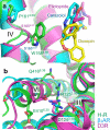
Fig. 2. Comparison of the structures of H1R, β2AR and D3R
(a) Proline-induced kink in helix IV (H1R: green, β2AR: cyan, D3R: magenta). The side chain of Trp1584.56 and Pro1614.59 of H1R and the equivalent residues of β2AR (Ser1654.57 and Pro1684.60) and of D3R (Ser1654.57 and Pro1674.59) are also shown. (b) Variations in the D(E)RY motif structures of H1R, β2AR and D3R colored in green, cyan and magenta, respectively. Side chains of Asp1243.49, Arg1253.50, Glu4106.30 of H1R and the equivalents of β2AR and D3R are represented as stick models. For H1R, Gln4166.36, which forms a hydrogen bond with Arg1253.50 , are also shown. Possible hydrogen bonds are indicated by dotted lines.
Fig. 3
Fig. 3. Binding interactions of doxepin
(a) Doxepin is shown as sticks with yellow carbon atoms, whereas the contact residues within 4 Å are shown with grey carbon atoms. Nitrogen and oxygen atoms are colored blue and red, respectively. Hydrogen bonds/salt bridges are indicated as blue dotted lines. (b) Doxepin binding interactions. Hydrophobic interactions are shown in black dotted lines. (c) Ligand binding positions in non-rhodopsin GPCRs. Carbon atoms of doxepin (H1R) are shown in yellow, carazolol (β2AR) in cyan, eticlopride (D3R) in magenta and ZM241385 (A2AAR) in grey. (d) Structure of the anion-binding region with a phosphate ion.
Fig.4
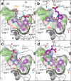
Fig.4. Interactions of second-generation selective H1R-antagonists with the H1R ligand-binding pocket
Conformation of each complex was predicted by global optimization of the ligand in the all-atom flexible H1R model ,, based on the H1R-doxepin complex structure. Carbon atoms for (a) Z-olopatadine co-bound with the phosphate ion, (b) acrivastine, (c) R-cetirizine (levocetirizine), and (d) fexofenadine are colored magenta. Nitrogen and oxygen atoms are colored blue and red, respectively. Ligand contact residues of H1R are shown with grey carbon atoms; parts of helices III, IV and ECL2 are not displayed for clarity. Hydrogen bonds are shown in cyan.

References

    1. Schwartz JC, Arrang JM, Garbarg M, Pollard H, Ruat M. Histaminergic transmission in the mammalian brain. Physiol Rev. 1991;71:1–51. - PubMed
    1. Hill SJ. Distribution, properties, and functional characteristics of three classes of histamine receptor. Pharmacol Rev. 1990;42:45–83. - PubMed
    1. Hill SJ, et al. International Union of Pharmacology. XIII. Classification of histamine receptors. Pharmacol Rev. 1997;49:253–278. - PubMed
    1. Yamashita M, et al. Expression cloning of a cDNA encoding the bovine histamine H1 receptor. Proc Natl Acad Sci U S A. 1991;88:11515–11519. - PMC - PubMed
    1. Simons FE. Advances in H1-antihistamines. N Engl J Med. 2004;351:2203–2217. - PubMed

Publication types

MeSH terms

Associated data