Review
. 2011 Jun 23;12(7):413-26.
doi: 10.1038/nrm3141.

The 'ins' and 'outs' of podosomes and invadopodia: characteristics, formation and function

Affiliations
Review

The 'ins' and 'outs' of podosomes and invadopodia: characteristics, formation and function

Danielle A Murphy et al. Nat Rev Mol Cell Biol. .

Abstract

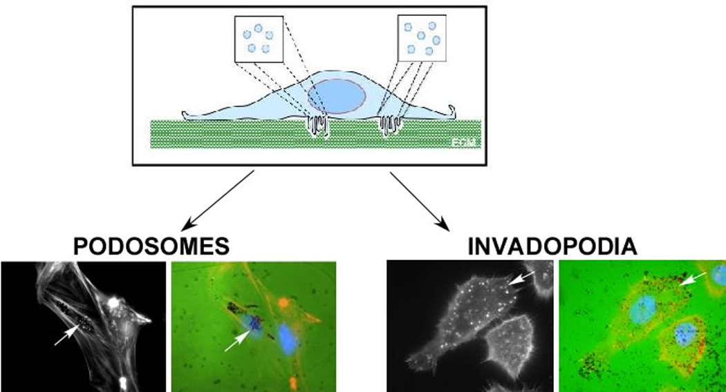
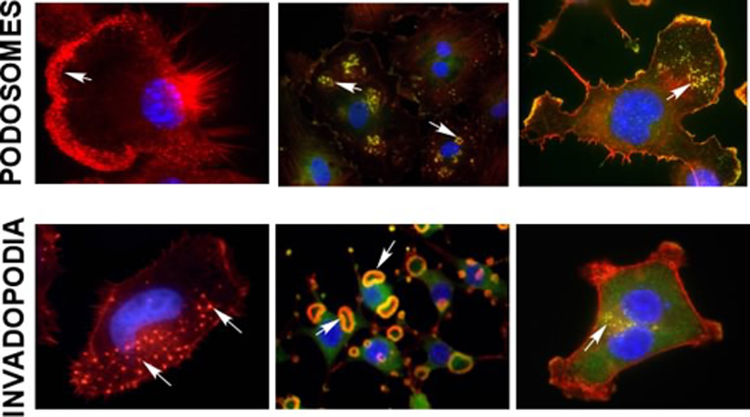
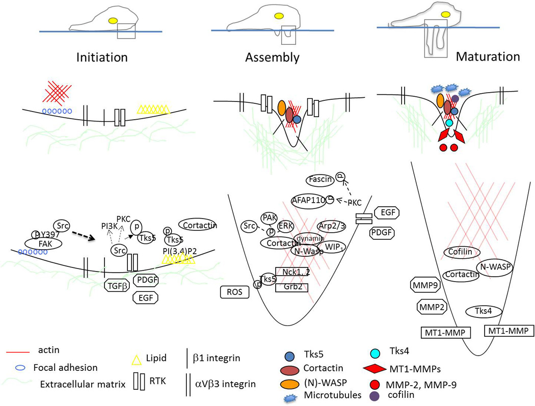
Podosomes and invadopodia are actin-based dynamic protrusions of the plasma membrane of metazoan cells that represent sites of attachment to - and degradation of - the extracellular matrix. The key proteins in these structures include the actin regulators cortactin and neural Wiskott-Aldrich syndrome protein (N-WASP), the adaptor proteins Tyr kinase substrate with four SH3 domains (TKS4) and Tyr kinase substrate with five SH3 domains (TKS5), and the metalloprotease membrane type 1 matrix metalloprotease (MT1MMP; also known as MMP14). Many cell types can produce these structures, including invasive cancer cells, vascular smooth muscle and endothelial cells, and immune cells such as macrophages and dendritic cells. Recently, progress has been made in our understanding of the regulatory and functional aspects of podosome and invadopodium biology and their role in human disease.


Figures

Figure 1
Figure 1. Regulators of Podosome and Invadopodia Formation
There are several components within the cell that are regulated to induce the formation and promote the function of podosomes and invadopodia. Some of the main components that are required for these structures are highlighted are, however this is not intended to be a totally comprehensive list.
Figure 2
Figure 2. Structure and Function of Podosomes and Invadopodia
Podosomes and invadopodia are actin rich structures that are formed on the ventral membrane of the cell. These structures are often seen as individual puncta or rosettes that protrude into the extracellular matrix (ECM). Classically, presence of these structures is often confirmed by culturing cells on top of fluorescently-conjugated matrix (FITC-gelatin), staining cells for F-actin and examining co-localization between F-actin puncta and degradation of matrix (black regions). This is demonstrated in vascular smooth muscle cells (podosomes) and SCC61 head and neck squamous carcinoma cells (invadopodia) as indicated by arrows.
Figure 3
Figure 3. Podosomes and Invadopodia in 2-dimensions
Formation of podosomes and invadopodia is frequently visualized by co-staining cells with F-actin (red) and the podosome and invadopodia associated protein cortactin (green). These structures can be seen in many cell types. Podosomes (top row): Macrophages (IC-21), vascular smooth muscle cells (VSMCs - A7r5 treated with 25nM PDGF) and neural crest stem cells (JOMA1.3, treated with 20nM PMA). Invadopodia (bottom row): head and neck squamous carcinoma cells (SCC61), Src-3T3 cells, and breast cancer cells (MDA-MB-231). Arrows denote podosomes and invadopodia.
Figure 4
Figure 4. Podosomes in vivo
Podosome structures have recently been observed in vivo. (A) Immunoelectron microscopy for Tks5 (red arrowhead) and cortactin (yellow arrowhead) in murine VSMCs in vivo. Enlarged image of labeled aorta section (boxed area, right panel) (B) Protrusive structures visible in migratory trunk neural crest cells in (foxd3:GFP) zebrafish, Danio rerio.
Figure 5
Figure 5. The stages of invadopodia formation
Initiation - Cells establish focal adhesions with the ECM through interaction of integrins, Src, and FAK (left side). In migrating cells, a switch occurs that will release Src to bind Tks5 and localize to regions containing PI(3,4)P2. These intracellular changes are initiated by factors such as EGF, PDGF, TGFβ (right side). Assembly - Formation of invadopodia occurs through recruitment and activation of actin regulatory proteins (Arp2/3, WIP), phosphorylation of key invadopodia components (cortactin, Tks5, fascin, AFAP110), and production of ROS. Maturation - Invadopodia promote degradation of ECM by coordinating secretion of MMP-2 and MMP-9, and enabling delivery (potentially through microtubules) and presentation of MT1-MMP to the tip of the protruding structure through the interaction of key invadopodia components (cortactin, Tks4, β1 integrin).

References

    1. David-Pfeuty T, Singer SJ. Altered distributions of the cytoskeletal proteins vinculin and alpha-actinin in cultured fibroblasts transformed by Rous sarcoma virus. Proc Natl Acad Sci U S A. 1980;77:6687–6691. - PMC - PubMed
    1. Tarone G, Cirillo D, Giancotti FG, Comoglio PM, Marchisio PC. Rous sarcoma virus-transformed fibroblasts adhere primarily at discrete protrusions of the ventral membrane called podosomes. Exp Cell Res. 1985;159:141–157. - PubMed
    1. Chen WT, Chen JM, Parsons SJ, Parsons JT. Local degradation of fibronectin at sites of expression of the transforming gene product pp60src. Nature. 1985;316:156–158. - PubMed
    1. Chen WT. Proteolytic activity of specialized surface protrusions formed at rosette contact sites of transformed cells. J Exp Zool. 1989;251:167–185. - PubMed
    1. Zambonin-Zallone A, Teti A, Carano A, Marchisio PC. The distribution of podosomes in osteoclasts cultured on bone laminae: effect of retinol. J Bone Miner Res. 1988;3:517–523. - PubMed

Publication types