

Planktonic microbes in the Gulf of Maine area
- PMID: 21698243
- PMCID: PMC3115965
- DOI: 10.1371/journal.pone.0020981
Planktonic microbes in the Gulf of Maine area
Abstract
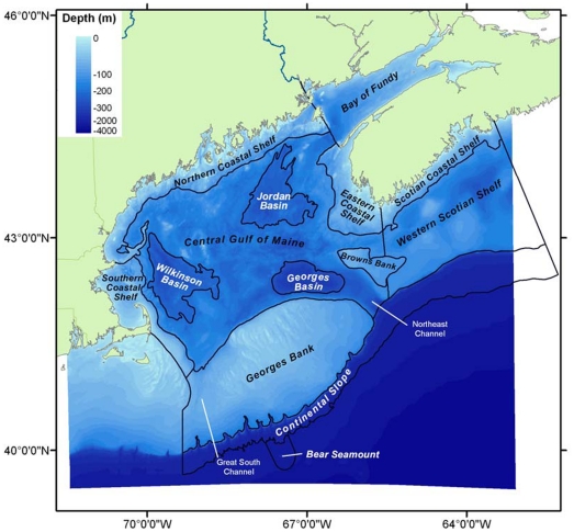
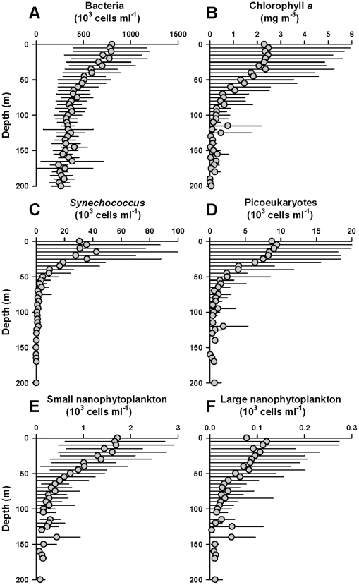
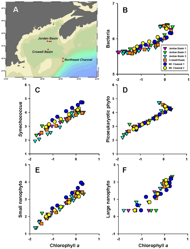
In the Gulf of Maine area (GoMA), as elsewhere in the ocean, the organisms of greatest numerical abundance are microbes. Viruses in GoMA are largely cyanophages and bacteriophages, including podoviruses which lack tails. There is also evidence of Mimivirus and Chlorovirus in the metagenome. Bacteria in GoMA comprise the dominant SAR11 phylotype cluster, and other abundant phylotypes such as SAR86-like cluster, SAR116-like cluster, Roseobacter, Rhodospirillaceae, Acidomicrobidae, Flavobacteriales, Cytophaga, and unclassified Alphaproteobacteria and Gammaproteobacteria clusters. Bacterial epibionts of the dinoflagellate Alexandrium fundyense include Rhodobacteraceae, Flavobacteriaceae, Cytophaga spp., Sulfitobacter spp., Sphingomonas spp., and unclassified Bacteroidetes. Phototrophic prokaryotes in GoMA include cyanobacteria that contain chlorophyll (mainly Synechococcus), aerobic anoxygenic phototrophs that contain bacteriochlorophyll, and bacteria that contain proteorhodopsin. Eukaryotic microalgae in GoMA include Bacillariophyceae, Dinophyceae, Prymnesiophyceae, Prasinophyceae, Trebouxiophyceae, Cryptophyceae, Dictyochophyceae, Chrysophyceae, Eustigmatophyceae, Pelagophyceae, Synurophyceae, and Xanthophyceae. There are no records of Bolidophyceae, Aurearenophyceae, Raphidophyceae, and Synchromophyceae in GoMA. In total, there are records for 665 names and 229 genera of microalgae. Heterotrophic eukaryotic protists in GoMA include Dinophyceae, Alveolata, Apicomplexa, amoeboid organisms, Labrynthulida, and heterotrophic marine stramenopiles (MAST). Ciliates include Strombidium, Lohmaniella, Tontonia, Strobilidium, Strombidinopsis and the mixotrophs Laboea strobila and Myrionecta rubrum (ex Mesodinium rubra). An inventory of selected microbial groups in each of 14 physiographic regions in GoMA is made by combining information on the depth-dependent variation of cell density and the depth-dependent variation of water volume. Across the entire GoMA, an estimate for the minimum abundance of cell-based microbes is 1.7×10(25) organisms. By one account, this number of microbes implies a richness of 10(5) to 10(6) taxa in the entire water volume of GoMA. Morphological diversity in microplankton is well-described but the true extent of taxonomic diversity, especially in the femtoplankton, picoplankton and nanoplankton--whether autotrophic, heterotrophic, or mixotrophic, is unknown.
Conflict of interest statement
Figures
References
-
- Snelgrove PVR. Cambridge University Press; 2010. Discoveries of the Census of Marine Life.
-
- Mills EL. New York: Cornell University Press; 1989. Biological oceanography: An early history, 1870-1960.
-
- Zwanenburg KCT, Bundy A, Strain P, Bowen WD, Breeze H, et al. Implications of ecosystem dynamics for the integrated management of the eastern Scotian Shelf. Can Tech Rep Fish Aquat Sci. 2006;2652:xiii +91.
Publication types
MeSH terms
LinkOut - more resources
Full Text Sources
Miscellaneous
