. 2011;6(6):e20806.
doi: 10.1371/journal.pone.0020806. Epub 2011 Jun 15.

Adenylate kinase 3 sensitizes cells to cigarette smoke condensate vapor induced cisplatin resistance

Affiliations

Adenylate kinase 3 sensitizes cells to cigarette smoke condensate vapor induced cisplatin resistance

Xiaofei Chang et al. PLoS One. 2011.

Abstract

Background: The major established etiologic risk factor for bladder cancer is cigarette smoking and one of the major antineoplastic agents used for the treatment of advanced bladder cancer is cisplatin. A number of reports have suggested that cancer patients who smoke while receiving treatment have lower rates of response and decreased efficacy of cancer therapies.

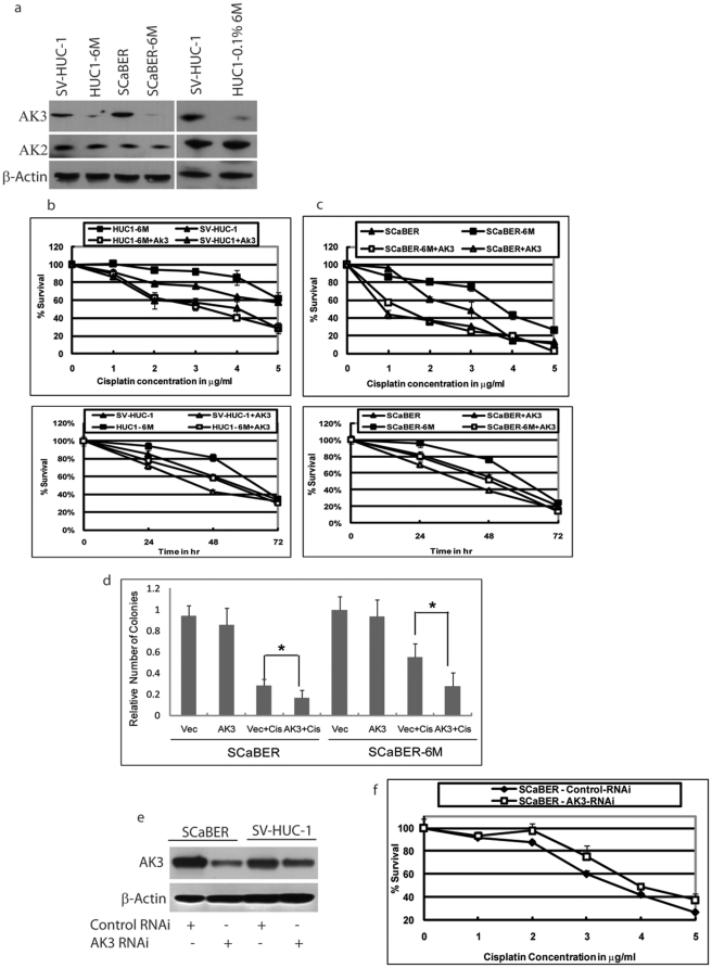
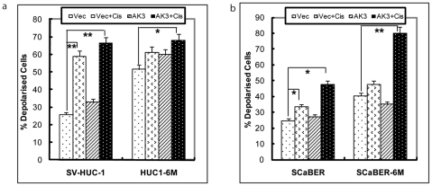
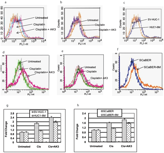
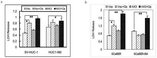
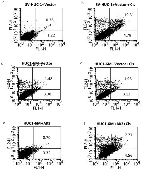
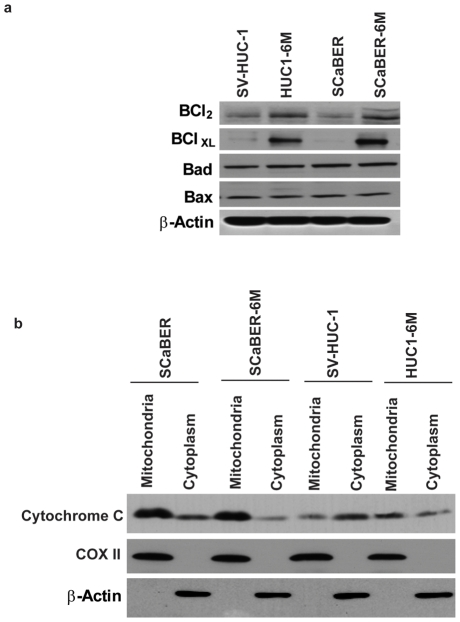
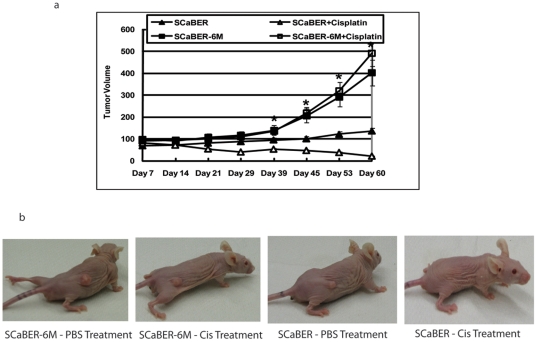
Methodology/principal findings: In this study, we investigated the effect of cigarette smoke condensate (CSC) vapor on cisplatin toxicity in urothelial cell lines SV-HUC-1 and SCaBER cells. We showed that chronic exposure to CSC vapor induced cisplatin resistance in both cell lines. In addition, we found that the expression of mitochondrial-resident protein adenylate kinase-3 (AK3) is decreased by CSC vapor. We further observed that chronic CSC vapor-exposed cells displayed decreased cellular sensitivity to cisplatin, decreased mitochondrial membrane potential (ΔΨm) and increased basal cellular ROS levels compared to unexposed cells. Re-expression of AK3 in CSC vapor-exposed cells restored cellular sensitivity to cisplatin. Finally, CSC vapor increased the growth of the tumors and also curtail the response of tumor cells to cisplatin chemotherapy in vivo.

Conclusions/significance: The current study provides evidence that chronic CSC vapor exposure affects AK3 expression and renders the cells resistant to cisplatin.


Conflict of interest statement

Competing Interests: The authors have declared that no competing interests exist.

Figures

Figure 1
Figure 1. CSC vapor decreased the expression of AK3 and increased cisplatin resistance.
(a) Western blot analysis was performed using anti-AK3 and anti-AK2 antiserum. Protein extracts in each lane are as indicated. Even loading was confirmed by re-probing the membrane with β-Actin antibody. HUC1-6M or SCaBER-6M indicates the cells chronically exposed to CSC vapor. HUC1-0.1%6M are SV-HUC-1 cells exposed to 0.1% CSC for 6 months. SV-HUC-1 and HUC1-6M (b), SCaBER and SCaBER-6M (c) were treated with 0 to 5 µg/ml cisplatin for 48 h, or with 3 µg/ml cisplatin for 24, 48 and 72 h, in the presence and absence of AK3 as indicated. (d) Colony formation assays were performed with SCaBER and SCaBER-6M in the presence and absence of AK3 as indicated. Data were expressed as mean ±SD. * indicates P<0.05 (e) SV-HUC-1 and SCaBER cells were transfected with AK3 RNAi for 48 h and Western blot analysis was performed using anti-AK3 antiserum. (f) SCaBER cells were transfected with AK3 RNAi and 48 h after transfection the cells were treated with 0 to 5 µg/ml cisplatin for 48 h and cellular survival was assessed. Error bars represent standard deviation of three experiments.
Figure 2
Figure 2. ΔΨm Alteration by AK3 in the Presence of Cisplatin.
Mitochondrial membrane potential was measured in SV-HUC1, HUC1-6M (a), SCaBER and SCaBER-6M (b) cells transfected with AK3 or control vector, followed by treatment with cisplatin for 48 h. Cells were stained with JC-1 reagent and cell fluorescence was measured on a flow cytometer using Fl1 and Fl2 channels. Increase of red fluorescence indicates hyper-polarization of mitochondrial membrane potential (ÄΨm). * indicates P<0.05 and ** indicates P<0.01.
Figure 3
Figure 3. ROS Production by AK3 in the Presence of Cisplatin.
ROS production was measured in parental SV-HUC-1 and SCaBER cells or cells exposed to CSC vapor for 6 months using DCFH-DA staining. The cells were transfected with or without the AK3 expression plasmid and treated with or without cisplatin for 48 h before staining. ROS generation in SV-HUC-1 and HUC1-6M (a–c) and SCaBER and SCaBER-6M (d–f) cells with overexpression of AK3 with and without cisplatin. The mean fluorescence density was calculated and data was plotted as mean ± SD (g,h).
Figure 4
Figure 4. AK3 Induced Release of Lactate Dehyderogenase upon Treatment of Cisplatin.
LDH release in SV-HUC-1 (a) and SCaBER (b) cells exposed to CSC vapor in presence of AK3 followed by cisplatin treatment for 48 h. The data are presented as means ± SD. All experiments were performed in triplicate. Statistical significance is as indicated with * indicates P<0.05 and ** indicates P<0.01
Figure 5
Figure 5. Activation of Apoptosis in the Presence of AK3 and Cisplatin.
Apoptosis was measured in the SV-HUC-1 and HUC1-6M cells using annexin/PI staining. The cells were transfected with or without AK3 expression plasmid and treated with or without cisplatin for 48 h as indicated.
Figure 6
Figure 6. CSC vapor induces Survival Proteins.
(a) Cellular lysates were obtained from the indicated cell lines and subjected to Western Blot analysis using the indicated antibodies. β-actin was used as a control. (b) The indicated cell lines were treated with 3 µg/ml cisplatin for 48 h. Mitochondrial and cytoplasmic total cellular lysates were subjected to Western Blot analysis using anti-cytochrome c antibody. Protein extracts in each lane are as indicated. Immunodetection of â-actin and COX II were done to assure that the transfected proteins were in cytoplasm and mitochondria respectively.
Figure 7
Figure 7. CSC Vapor Induces Cisplatin Resistance In Xenograft Model.
(a) Subcutaneous xenografts were generated in of 5- to 6-week-old female athymic nu/nu mice with SCaBER or SCaBER-6M cells. One week following tumor cell inoculation, mice were treated with either PBS or cisplatin (3 mg/Kg/dose). The mean tumor volume was calculated. * indicates P<0.01 (b) One representative mouse from each group was displayed.

References

    1. Jankovic S, Radosavljevic V. Risk factors for bladder cancer. Tumori. 2007;93:4–12. - PubMed
    1. Hecht SS. Tobacco smoke carcinogens and breast cancer. Environ Mol Mutagen. 2002;39:119–126. - PubMed
    1. Skipper PL, Tannenbaum SR, Ross RK, Yu MC. Nonsmoking-related arylamine exposure and bladder cancer risk. Cancer Epidemiol Biomarkers Prev. 2003;12:503–507. - PubMed
    1. Boffetta P. Epidemiology of environmental and occupational cancer. Oncogene. 2004;23:6392–6403. - PubMed
    1. Barsanti KC, Luo W, Isabelle LM, Pankow JF, Peyton DH. Tobacco smoke particulate matter chemistry by NMR. Magn Reson Chem. 2007;45:167–170. - PubMed

Publication types