

The clinical relevance of bifid and trifid mandibular canals
- PMID: 21698363
- PMCID: PMC3294215
- DOI: 10.1007/s10006-011-0278-5
The clinical relevance of bifid and trifid mandibular canals
Abstract
Background: Bifid mandibular canals (BMC) and trifid mandibular canals (TMC) are variations on the normal anatomy with incidences ranging from 0.08% to 65.0%. Such aberrations have an important clinical impact. For example, an extra mandibular canal may explain inadequate anesthesia, especially when two mandibular foramina are involved. Furthermore, during mandibular surgery, a second, or even third, neurovascular bundle may be damaged causing paresthesia, neuroma development, or bleeding.
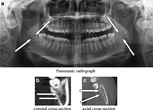
Case report: Two cases are presented in this article. One patient had a BMC on both sites, and the other patient had a TMC on one site and a BMC on the other site.
Discussion: Initial screening for the presence of a BMC or TMC can be executed by conventional panoramic radiography. BMCs or TMCs are diagnosed, before executing mandibular surgery; additional CBCT scanning is indicated.
Figures


References
-
- Anderson LC, Kosinski TF, Mentag PJ. A review of the intraosseous course of the nerves of the mandible. J Oral Implantol. 1991;17:394–403. - PubMed
-
- Auluck A, Pai KM, Mupparapu M. Multiple mandibular nerve canals: radiographic observations and clinical relevance. Report of 6 cases. Quintessence Int. 2007;38:781–787. - PubMed
Publication types
MeSH terms
LinkOut - more resources
Full Text Sources
