

The relationships among spatiotemporal collagen gene expression, histology, and biomechanics following full-length injury in the murine patellar tendon
- PMID: 21698662
- PMCID: PMC3181390
- DOI: 10.1002/jor.21484
The relationships among spatiotemporal collagen gene expression, histology, and biomechanics following full-length injury in the murine patellar tendon
Abstract
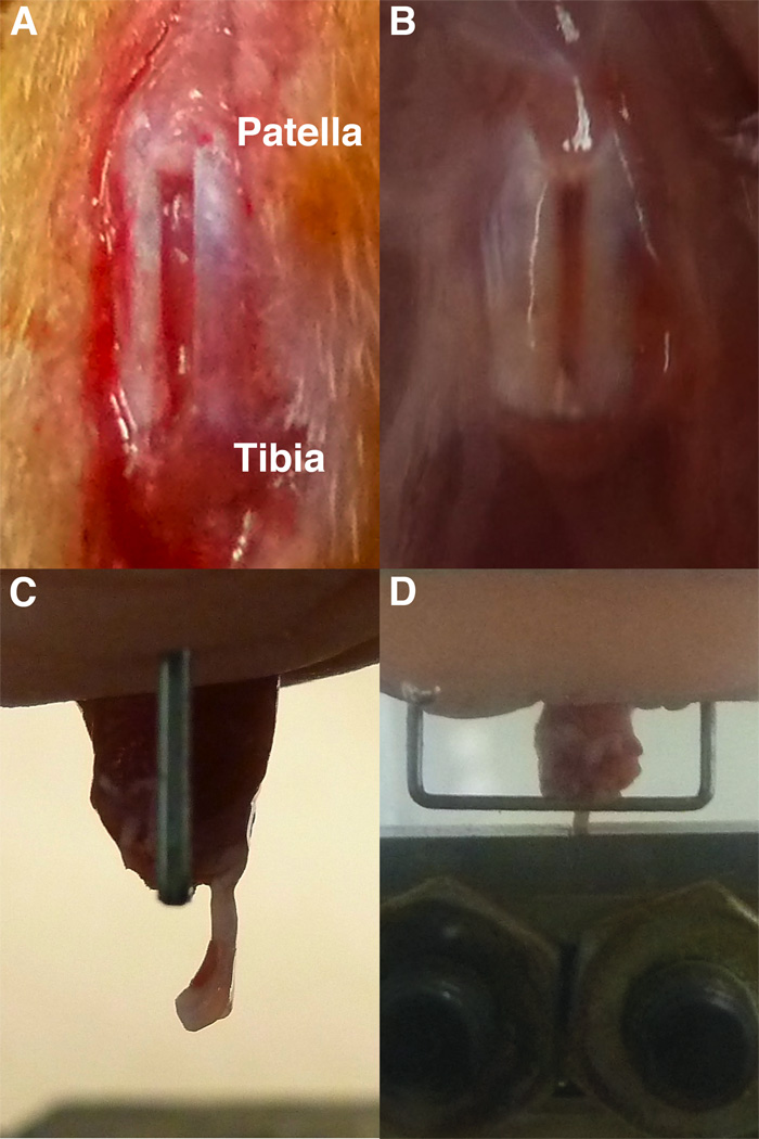
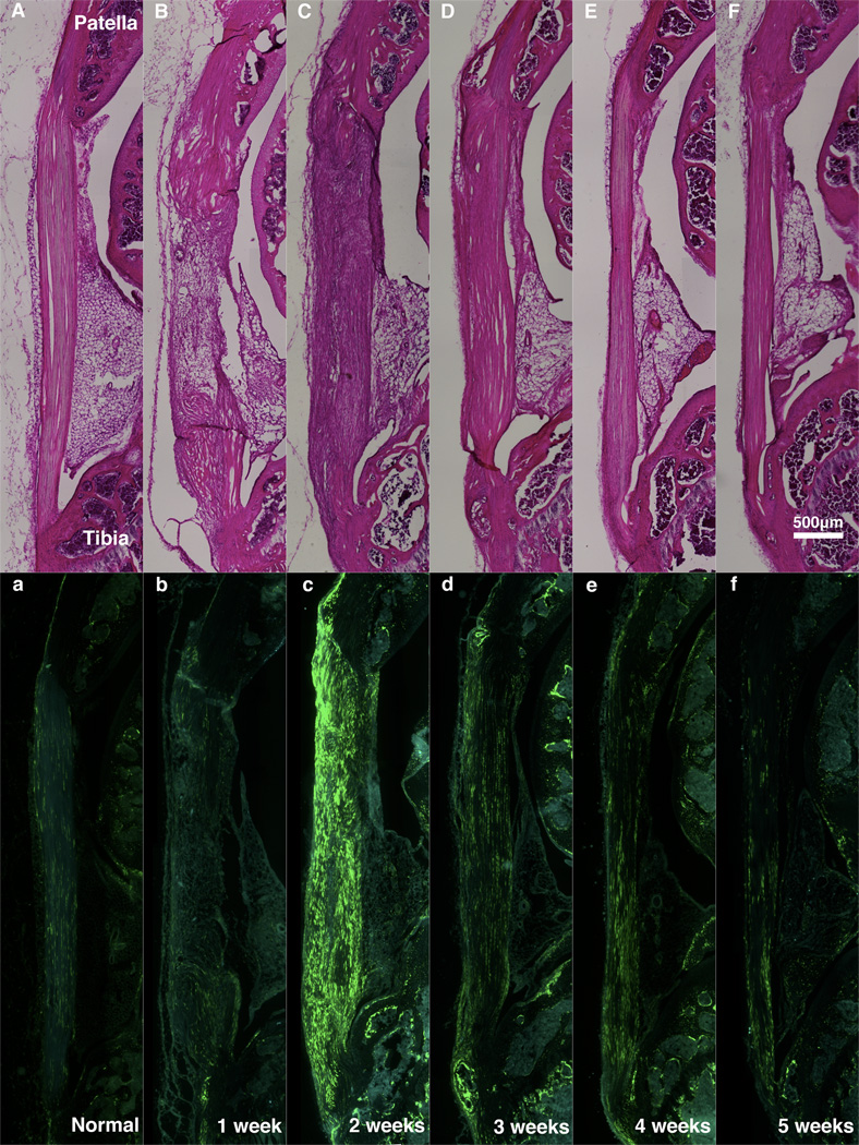
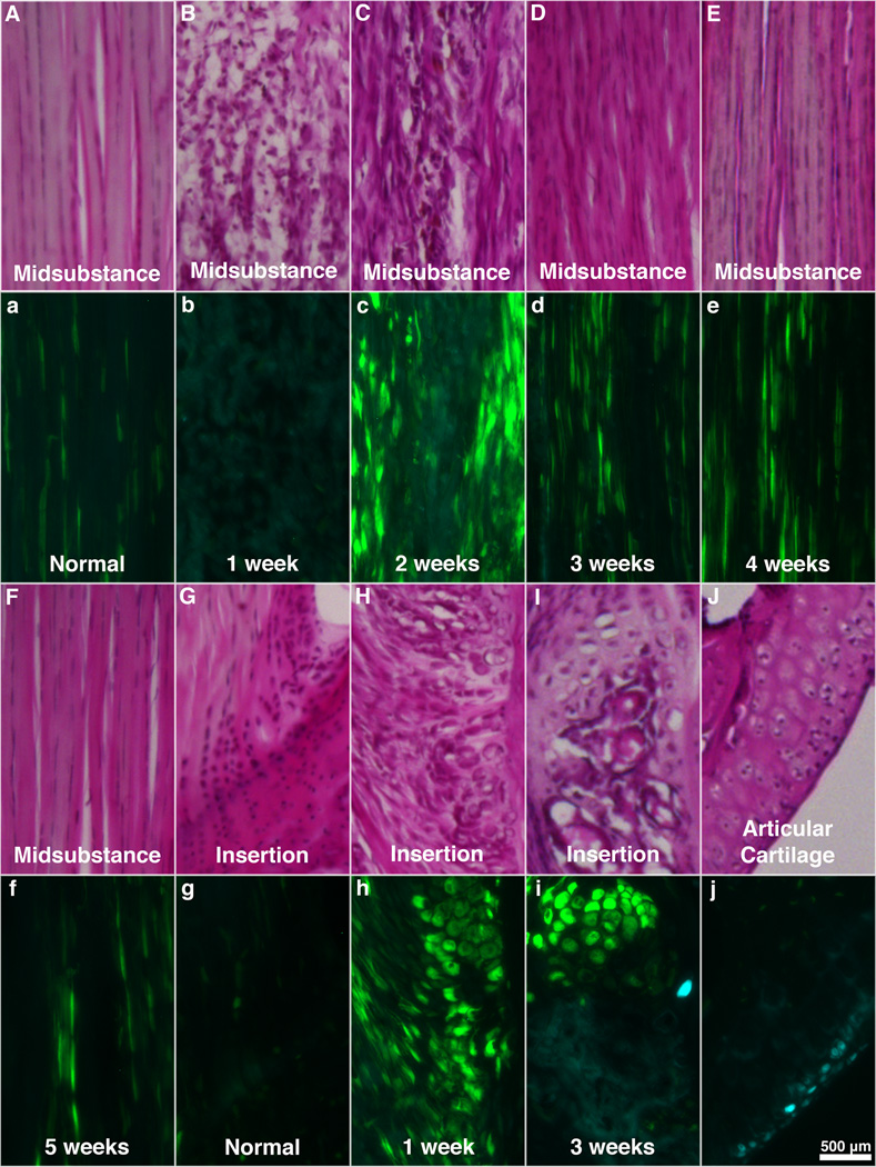
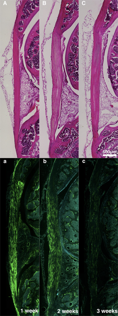
Tendon injuries are major orthopedic problems that worsen as the population ages. Type-I (Col1) and type-II (Col2) collagens play important roles in tendon midsubstance and tendon-to-bone insertion healing, respectively. Using double transgenic mice, this study aims to spatiotemporally monitor Col1 and Col2 gene expression, histology, and biomechanics up to 8 weeks following a full-length patellar tendon injury. Gene expression and histology were analyzed weekly for up to 5 weeks while mechanical properties were measured at 1, 2, 5, and 8 weeks. At week 1, the healing region displayed loose granulation tissue with little Col1 expression. Col1 expression peaked at 2 weeks, but the ECM was highly disorganized and hypercellular. By 3 weeks, Col1 expression had reduced and by 5 weeks, the ECM was generally aligned along the tendon axis. Col2 expression was not seen in the healing midsubstance or insertion at any time point. The biomechanics of the healing tissue was inadequate at all time points, achieving ultimate loads and stiffnesses of 48% and 63% of normal values by 8 weeks. Future studies will further characterize the cells within the healing midsubstance and insertion using tenogenic markers and compare these results to those of tendon cells during normal development.
Copyright © 2011 Orthopaedic Research Society.
Figures
References
-
- Praemer A, Furner S, Rice D. Musculoskeletal conditions in the United States. Second ed. Parke Ridge, IL: American Academy of Orthopaedic Surgeons; 1999.
-
- Rees JD, Wilson AM, Wolman RL. Current concepts in the management of tendon disorders. Rheumatology. 2006;45:508–521. - PubMed
-
- Thomopoulos S, Marquez J, Weinberger B, et al. Collagen fiber orientation at the tendon to bone insertion and its influence on stress concentrations. J Biomech. 2006;39:1842–1851. - PubMed
-
- Galatz L, Rothermich S, VanderPloeg K, et al. Development of the supraspinatus tendon-to-bone insertion: Localized expression of extracellular matrix and growth factor genes. J Orthop Res. 2007;25:1621–1628. - PubMed
Publication types
MeSH terms
Substances
Grants and funding
LinkOut - more resources
Full Text Sources
Medical
Molecular Biology Databases
