Review
. 2011 Nov 1;15(9):2477-86.
doi: 10.1089/ars.2011.3976.

Control of reactive oxygen species production in contracting skeletal muscle

Affiliations
Review

Control of reactive oxygen species production in contracting skeletal muscle

Malcolm J Jackson. Antioxid Redox Signal. .

Abstract

Significance: The increased activities of free radicals or reactive oxygen species in tissues of exercising humans and animals were first reported ∼30 years ago. A great deal has been learned about the processes that can generate these molecules, but there is little agreement on which are important, how they are controlled, and there are virtually no quantitative data. Superoxide and nitric oxide are generated by skeletal muscle and their reactions lead to formation of secondary species. A considerable amount is known about control of superoxide generation by xanthine oxidase activity, but similar information for other generation systems is lacking.

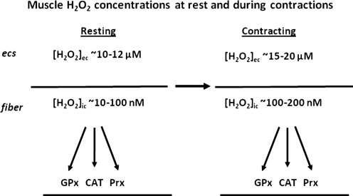
Recent advances: Re-evaluation of published data indicates potential approaches to quantification of the hydrogen peroxide concentration in resting and contracting muscle cells. Such calculations reveal that, during contractions, intracellular hydrogen peroxide concentrations in skeletal muscle may only increase by ∼100 nM. The primary effects of this modest increase appear to be in "redox" signaling processes that mediate some of the responses and adaptations of muscle to exercise. These act, in part, to increase the expression of cytoprotective proteins (e.g., heat shock proteins and antioxidant enzymes) that help maintain cell viability. During aging, these redox-mediated adaptations fail and this contributes to age-related loss of skeletal muscle.

Critical issues and future directions: Understanding the control of ROS generation in muscle and the effect of aging and some disease states will aid design of interventions to maintain muscle mass and function, but is dependent upon development of new analytical approaches. The final part of this review indicates areas where such developments are occurring.


Figures

FIG. 1.
FIG. 1.
Schematic representation of the sites and mechanisms proposed for ROS and NO generation in skeletal muscle fibers. Scheme updated from Powers and Jackson (60) to incorporate new data on the potential for hydrogen peroxide release from fibers to the extracellular space (see text for details) and on the role of CuZnSOD in the mitochondrial intermembrane space (IMS). (To see this illustration in color the reader is referred to the web version of this article at www.liebertonline.com/ars).
FIG. 2.
FIG. 2.
Comparison of the rate of increase in CM-DCF fluorescence from single isolated fibers from mouse flexor digitorum brevis muscles subjected to either a 15 min period of electrically stimulated isometric contractions (A) or exposed to 1 μM hydrogen peroxide for 15 min (B). The increase in CM-DCF fluorescence from contracting fibers was less than 50% of that seen following exposure to hydrogen peroxide. Data redrawn from Palomero et al, (57).
FIG. 3.
FIG. 3.
The concentration of hydrogen peroxide in microdialysates from the gastrocnemius muscles of mice over five 15 min collections at rest, followed by 15 min of isometric contractions and a further 15 min at rest (A). A schematic diagram of the microdialysis probe is also shown (B), together with the calculation of interstitial hydrogen peroxide from microdialysate values and the values for the recovery of hydrogen peroxide across the probe obtained in preliminary studies. Data derived from Vasilaki et al. (76).
FIG. 4.
FIG. 4.
Schematic representation of the concentrations of hydrogen peroxide calculated to occur in the cytosol of muscle fibers ([H2O2]ic) and interstitial space ([H2O2]ec)of fibers at rest and following the period of 15 min contractions. See text for details of calculations. CAT, catalase; ECS, extracellular space; GPx, glutathione peroxidase; Prx: peroxiredoxins.
FIG. 5.
FIG. 5.
Comparison of the effects of contraction on superoxide release from gastrocnemius muscles of old (black bars) compared with muscles of adult mice (open bars). Data are from microdialysis studies and show results from 15 min collections at rest, followed by 15 min of isometric contractions. *P<0.05 compared with the superoxide content in microdialysate from adult mice prior to contractions. Data derived from Vasilaki et al. (76).
FIG. 6.
FIG. 6.
Images of single isolated fibers from mouse FDB muscles loaded with ROS sensitive probes. (A) Brightfield image, bar=50 micron; (B) fiber loaded with DHE; (C) the same fiber as in B stained with DAPI nuclear stain; (D) fiber loaded with DCFH. (To see this illustration in color the reader is referred to the web version of this article at www.liebertonline.com/ars).

References

    1. Antunes F. Cadenas E. Cellular titration of apoptosis with steady state concentrations of H(2)O(2): Submicromolar levels of H(2)O(2) induce apoptosis through Fenton chemistry independent of the cellular thiol state. Free Radic Biol Med. 2001;30:1008–1018. - PubMed
    1. Arbogast S. Reid MB. Oxidant activity in skeletal muscle fibers is influenced by temperature, CO2 level, and muscle-derived nitric oxide. Am J Physiol Regul Integr Comp Physiol. 2004;287:R698–705. - PubMed
    1. Arora S. Vaishya R. Dabla PK. Singh B. NAD(P)H oxidases in coronary artery disease. Adv Clin Chem 2010. 2010;50:65–86. - PubMed
    1. Bakkar N. Wang J. Ladner KJ. Wang H. Dahlman JM. Carathers M. Acharyya S. Rudnicki MA. Hollenbach AD. Guttridge DC. IKK/NF-kappaB regulates skeletal myogenesis via a signaling switch to inhibit differentiation and promote mitochondrial biogenesis. J Cell Biol. 2008;180:787–802. - PMC - PubMed
    1. Belousov VV. Fradkov AF. Lukyanov KA. Staroverov DB. Shakhbazov KS. Terskikh AV. Lukyanov S. Genetically encoded fluorescent indicator for intracellular hydrogen peroxide. Nat Methods. 2006;3:281–286. - PubMed

Substances

LinkOut - more resources