Review
. 2012 Mar;165(6):1617-1643.
doi: 10.1111/j.1476-5381.2011.01551.x.

Pharmacological modulation of chemokine receptor function

Affiliations
Review

Pharmacological modulation of chemokine receptor function

D J Scholten et al. Br J Pharmacol. 2012 Mar.

Abstract

G protein-coupled chemokine receptors and their peptidergic ligands are interesting therapeutic targets due to their involvement in various immune-related diseases, including rheumatoid arthritis, multiple sclerosis, inflammatory bowel disease, chronic obstructive pulmonary disease, HIV-1 infection and cancer. To tackle these diseases, a lot of effort has been focused on discovery and development of small-molecule chemokine receptor antagonists. This has been rewarded by the market approval of two novel chemokine receptor inhibitors, AMD3100 (CXCR4) and Maraviroc (CCR5) for stem cell mobilization and treatment of HIV-1 infection respectively. The recent GPCR crystal structures together with mutagenesis and pharmacological studies have aided in understanding how small-molecule ligands interact with chemokine receptors. Many of these ligands display behaviour deviating from simple competition and do not interact with the chemokine binding site, providing evidence for an allosteric mode of action. This review aims to give an overview of the evidence supporting modulation of this intriguing receptor family by a range of ligands, including small molecules, peptides and antibodies. Moreover, the computer-assisted modelling of chemokine receptor-ligand interactions is discussed in view of GPCR crystal structures. Finally, the implications of concepts such as functional selectivity and chemokine receptor dimerization are considered.


Figures

Figure 1
Figure 1
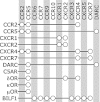
Overview of chemokine receptors (vertical) and their ligands (horizontal). An open circle indicates the specific ligand binds to the receptor connected to it by a black line. From top to bottom: human chemokine receptors (XCR1 to CX3CR1), chemokine decoy receptors (D6 to CCX CKR) and viral chemokine receptors (US28 to U51). For reasons of clarity, this figure does not show differences in affinity or activity between ligands for the same receptor. For more detailed information on ligand properties, visit the website of The International Union of Basic and Clinical Pharmacology (IUPHAR) (Murphy et al., 2011).
Figure 2
Figure 2
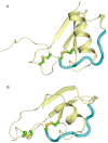
Overall tertiary structure of chemokines. (A) Structure of a CC-chemokine (CCL5). (B) Structure of a CXC-family chemokine (CXCL12). The disulfide bonds are indicated in green and yellow. The N-loop is shown in cyan. See text for more details.
Figure 3
Figure 3
Chemokine receptors, their cellular expression profile and association with disease. The cellular expression profiles of the chemokine receptors are shown by open circles on the left-hand side. The cells are divided in haematopoietic cells and miscellaneous cells. Meaning of abbreviations: VSMC, vascular smooth muscle cells; VE, vascular endothelial cells; LE, lymphatic endothelial cells; BSMC, bronchial smooth muscle cells. The association of a particular receptor with disease is shown on the right-hand side of the figure. The diseases are categorized into inflammatory diseases and cancer. Abbreviations: IBD, inflammatory bowel disease; COPD, chronic obstructive pulmonary disease.
Figure 4
Figure 4
Schematic overview of GPCRs, their activation and ligand-binding sites. (A) The overall topology of a GPCR structure, with annotated TM helices, and N- and C-terminus. (B,C) Schematic representation of the two-step model of chemokine receptor activation. (B) The first step: chemokine binding to the extracellular surface of the chemokine receptor, including the N-terminus (CRS1). (C) The second step: the flexible N-terminus of the chemokine is positioned to interact with EL and TM residues (CRS2), mediating receptor activation. (D) Intracellular allosteric binding pocket used by CXCR2 and CCR4 ligands. Also the G protein binds in this region. (E) Binding pocket for small-molecule ligands in the chemokine receptor between TM1, 2, 3 and 7 (TMS1). (F) Binding pocket for small-molecule ligands in chemokine receptors between TM3, 4, 5, 6 and 7 (TMS2). Table 1 and Figure 5 give an overview of the interaction residues of small ligands binding to chemokine receptors.
Figure 6
Figure 6
Crystal structures of CXCR4 receptors complexed with ligands. (A) Comparison of the ligand binding modes of the small-molecule antagonist IT1t (magenta, 3odu) and the peptide-like ligand CVX15 (green, 3oe0) in the (IT1t bound) CXCR4 crystal structure. While IT1t binds in the TMS1 (minor pocket) between TM helices 1, 2, 3 and 7, CVX15 binds primarily in TMS2 (major pocket) between TMs 3, 4, 5, 6 and 7. TMs 2, 4 and 7 are coloured cyan, yellow and orange respectively, and Cα atoms of residues D2.63 (TMS1), D4.60 (TMS2) and E7.39 (interface between TMS1 and TMS2) are indicated with black spheres (like in panels B, D and E). Furthermore, the beta sheet of EL2 and the disordered helix 8 are labelled. (B) Interactions between the residues 1–4 and 13–14 of CVX15 (green side chain in ball-and-stick, backbone as ribbon) and CXCR4 (3oe0). Important residues are displayed as ball-and-stick (grey carbon atoms), while CVX15-1T1t H-bond and ionic interactions are indicated with black and grey dashed lines respectively. Nitrogen, oxygen and sulphur atoms are coloured blue, red and yellow respectively. For reasons of clarity the top of TM3 is not shown, while only the first β strand of EL2 is displayed. (C) Chemical structures of IT1t and CVX15 (part of molecule displayed in panel B is coloured green). (D,E) Comparison of the interactions between IT1t (magenta carbon atoms) and CXCR4 in the experimentally determined X-ray crystal structure (3odu, panel D) and the best in silico CXCR4 model in the worldwide GPCR DOCK 2010 competition (panel E) correctly predicting the highest number of IT1t-CXCR4 contacts (prior to release of the CXCR4-IT1t crystal structure). Important residues are displayed as ball-and-stick (grey carbon atoms), while IT1t-CXCR4 H-bonds are indicated with black dashed lines. Colour coding of helices and heteroatoms are the same as defined in panels A and B. For reasons of clarity the top of TM3 is not shown.
Figure 5
Figure 5
Alignment of important amino acid residues of TM domains and EL2. The TM residues are shown using the Ballesteros–Weinstein (B&W) numbering scheme (Ballesteros and Weinstein, 1995). An adapted version is also used for the EL2 residues (45.50 and 45.51) indicating residues in the loop between TM4 and TM5, using the conserved cysteine as reference: 45.50 (de Graaf et al., 2008). Mutation of residues highlighted in gray significantly affected ligand binding. Also the TXP motif is shown (2.56–2.58), which is conserved among chemokine receptors (see text for details). The residues are coloured blue in case of TMS1 residues (minor pocket), orange for TMS2 residues (major pocket), yellow for interface residues between TMS1 and TMS2, and green for intracellular residues. For CXCR4, contacts from the crystal structures of the receptor with ligands are indicated with a thick black border. Magenta indicates contacts for the small-molecule IT1t, green for the peptide CVX15, and orange for both ligands. For more detailed information on the residues, ligands and associated literature references, see Table 1.
Figure 7
Figure 7
GPCR dimerization partners of chemokine receptors. The open circles connect the chemokine receptors with their suggested dimerization partners.

References

    1. Ahuja S, Lee J, Murphy P. CXC chemokines bind to unique sets of selectivity determinants that can function independently and are broadly distributed on multiple domains of human interleukin-8 receptor B. Determinants of high affinity binding and receptor activation are distinct. J Biol Chem. 1996;271:225–232. - PubMed
    1. Alexander SPH, Mathie A, Peters JA. Guide to Receptors and Channels (GRAC), 5th Edition. Br J Pharmacol. 2011;164(Suppl. 1):S1–S324. - PMC - PubMed
    1. Allegretti M, Bertini R, Cesta M, Bizzarri C, Di Bitondo R, Di Cioccio V, et al. 2-Arylpropionic CXC chemokine receptor 1 (CXCR1) ligands as novel noncompetitive CXCL8 inhibitors. J Med Chem. 2005;48:4312–4331. - PubMed
    1. Allen S, Crown S, Handel T. Chemokine: receptor structure, interactions, and antagonism. Annu Rev Immunol. 2007;25:787–820. - PubMed
    1. Andrews G, Jones C, Wreggett KA. An intracellular allosteric site for a specific class of antagonists of the CC chemokine G protein-coupled receptors CCR4 and CCR5. Mol Pharmacol. 2008;73:855–867. - PubMed

MeSH terms

LinkOut - more resources