Comparative Study
. 2011 Oct;20(5):587-98.
doi: 10.1111/j.1365-2583.2011.01089.x. Epub 2011 Jun 24.

Comparison of transgene expression in Aedes aegypti generated by mariner Mos1 transposition and ΦC31 site-directed recombination

Affiliations
Comparative Study

Comparison of transgene expression in Aedes aegypti generated by mariner Mos1 transposition and ΦC31 site-directed recombination

Alexander W E Franz et al. Insect Mol Biol. 2011 Oct.

Abstract

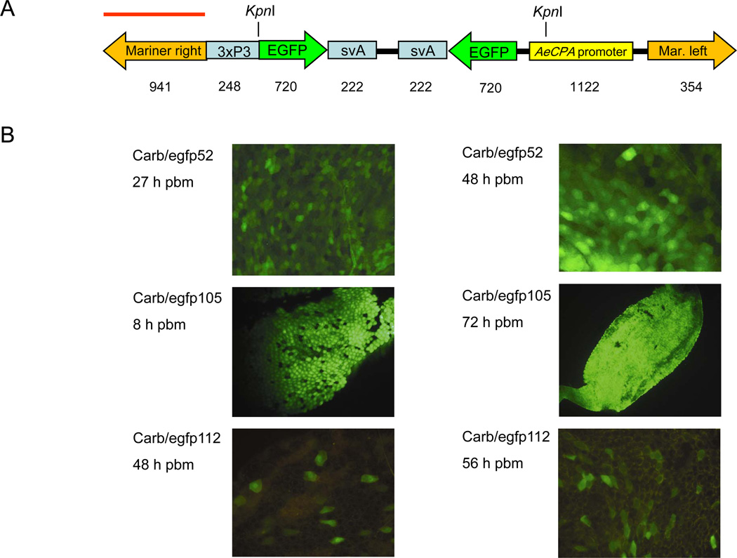
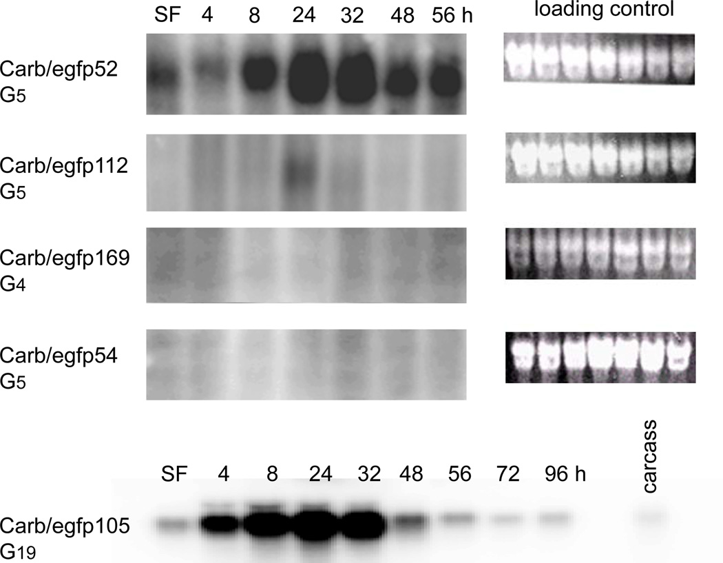
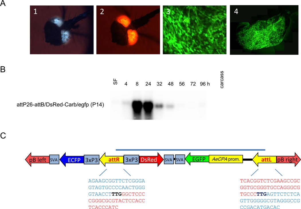
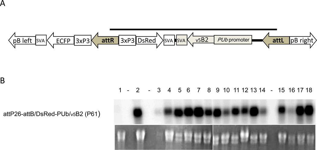
Transgenic mosquitoes generated by transposable elements (TEs) often poorly express transgenes owing to position effects. To avoid these effects, the ΦC31 site-directed recombination system was used to insert transgenes into a locus favourable for gene expression in Aedes aegypti. We describe phenotypes of mariner Mos1 TE and ΦC31 transgenic mosquitoes expressing the enhanced green fluorescent protein (EGFP) reporter in midguts of blood-fed females. Mosquitoes of nine TE-generated lines [estimated transformation frequency (TF): 9.3%] clearly expressed the eye-specific selection marker but only 2/9 lines robustly expressed the EGFP reporter. The piggyBac TE-generated ΦC31 docking strain, attP26, supported recombination with attB site containing donors at an estimated TF of 1.7-4.9%. Using a codon-optimized ΦC31 integrase mutant instead of the 'wild-type' enzyme did not affect TF. Site-directed recombination of line attP26 with an attB-containing donor expressing EGFP from the Ae. aegypti carboxypeptidase promoter produced one transgenic line with blood-fed females expressing the reporter in midgut tissue. Docking strain attP26 also supported robust expression of Flock House virus B2 from the Ae. aegypti polyubiquitin promoter. Our data confirm that eye-specific selection marker expression alone is not a reliable indicator for robust gene-of-interest expression in Ae. aegypti and that the ΦC31 system can ensure predictable transgene expression in this mosquito species.


Figures

Figure 1
Figure 1
Midgut-specific reporter gene expression in mariner Mos1 transformed Aedes aegypti. (A). Diagram of the mariner Mos1 TE based transgene pMos-carb/EGFP/svA. Locations of KpnI sites and probe (red bar) for Southern analysis are indicated. Numbers below the diagram indicate DNA fragment sizes in base pairs (bp). (B). EGFP expression in female midguts of the pMos-carb/EGFP/svA transformed lines Carb/egfp52, 105, and 112 at 8–72 h post-bloodmeal (pbm). EGFP was viewed under a fluorescent microscope equipped with EGFP specific filter sets.
Figure 2
Figure 2
Northern blot analyses of EGFP transcription in female midguts of the mariner Mos1 transformed lines (G4–5) Carb/egfp52, 112, 169, 54, and (G19) 105. Samples were prepared from midguts harvested after sugarfeeding (SF) or 4–96 h pbm. The 10 carcasses of Carb/egfp105 mosquitoes were without heads. The hybridization pattern of Carb/egfp157, 188, and 191 (not shown) resembled those of Carb/egfp54 and 169. Total RNA was extracted from pools of 25 midguts per time point. Blots were hybridized with random primed 32P-dCTP- labeled DNA probes or in vitro-transcribed 32P-dUTP-labeled probes corresponding to the EGFP gene at 48°C. Ethidium bromide stained agarose gels are shown as loading controls.
Figure 3
Figure 3
Genotypic analysis of transgenic mosquito lines. (A). Southern blot analysis of mariner Mos1 transformed lines Carb/egfp52, 54, 86, 105, and 112. Total DNA was extracted from 10 females per line, followed by digestion with KpnI. Blots were hybridized with a random-primed 32P-dCTP-labeled DNA probe corresponding to the right arm of the mariner Mos1 TE at 48°C. (B). Detection of the attP site-specific recombination site in transgenic mosquitoes of lines attP26 and attP35. Total DNA of five females was extracted and used as template for gene amplification using primers corresponding to the 171 bp attP site and 220 bp flanking cloning sites of plasmid Bac[3xP3-ECFPfa]attP shown above as a diagram (Nimmo et al., 2006). (C). Southern blot analysis of piggyBac transformed lines attP26 and attP35. Total DNA was extracted from 3 females per line, followed by digestion with EcoRV. Blots were hybridized with a random primed 32P-dCTP labeled DNA probe corresponding to the left arm/3xP3/ECFP encoding region of the piggyBac TE at 48°C. (D). Partial sequence (nt position 2,685,212–2,685,388) of the Ae. aegypti genomic DNA of supercontig 1.2, contig 1407. Highlighted in blue: partial sequence of piggyBac left arm; highlighted in green: partial sequence of piggyBac right arm. The CTAG sequence motif is underlined and/or in bold.
Figure 4
Figure 4
Codon optimization of the ΦC31 integrase and ΦC31-mediated recombination of line attP26 with the attB/DsRed2 donor plasmid. (A). Codon optimization was performed on the DNA sequence of ΦC31 integrase P3 mutant (Keravala et al., 2009), which contained an additional 33 amino acid sequence upstream of the ‘wild-type’ ΦC31 start codon. The DNA sequence of the P3 mutant was codon optimized according to the most frequent codon usage in the An. stephensi genome, which is similar to that of Ae. aegypti. (B). Diagram of the attB/DsRed2 donor plasmid integration into the genome of docking strain attP26 following recombination between attB and attP in presence of ΦC31(‘wild-type’) or codon optimized ΦC31 P3 mutant integrases, respectively. As a consequence of recombination, attP and attB sites are converted into attL and attR. Grey bar indicates the part of the transgene that originates from the donor plasmid.
Figure 5
Figure 5
ΦC31-mediated recombination of docking strain attP26 with the attB/DsRed2-carb/EGFP/svA donor plasmid. (A). Eye-specific marker gene expression in line P14. (1) ECFP expression originates from the selection marker of line attP26; (2) DsRed expression originates from the donor plasmid. (3) Midgut-specific EGFP expression represents the gene-of-interest of the donor and was visible at 48 h post-bloodmeal (pbm) in individual epithelial cells of (4) the entire midgut containing a bloodmeal. (B). Northern blot analysis for the detection of EGFP transcripts in female midguts of attP26-attB/DsRed-carb/egfp (P14) mosquitoes after sugarfeeding (SF) or 4–96 h pbm. Total RNA was extracted from 25 midguts per time point or 10 headless carcasses. Blots were hybridized with a random primed 32P-dCTP labeled DNA probe corresponding to the EGFP gene at 48°C. (C). Diagram of the donor plasmid integration pattern following recombination between the attP site of line attP26 and the attB site of the donor. attP and attB sites are converted into attL and attR consisting of DNA sequence originating from attP (blue) and attB (red). In bold and black: crossover recognition motif. attL and attR sequences were confirmed by gene amplification and DNA sequencing of the amplicons. Grey bar indicates the part of the transgene that originates from the donor plasmid.
Figure 6
Figure 6
ΦC31-mediated recombination of line attP26 with the attB/DsRed2-PUb/v5B2/ svA donor plasmid to constitutively express the Flock House virus B2 suppressor of RNA interference in Ae. aegypti. (A) Diagram of the donor plasmid integration pattern following recombination between the attP site of line attP26 and the attB site of the donor. Grey bar indicates the part of the transgene that originates from the donor plasmid. (B) Northern blot showing v5B2 expression over time in various tissues of recombinant P61 mosquitoes. Lanes: (1) docking strain attP26 (negative control); (2) P61 larvae; (3) P61 females - midguts, 1 day post-emergence (pe); (4) carcasses, 1 day pe; (5) midguts, 3 days pe; (6) carcasses, 3 days pe; (7) midguts, 5 days pe; (8) carcasses, 5 days pe; (9) midguts, 7 days pe; (10) carcasses, 7 days pe; (11) midguts of bloodfed females, 7 days post-bloodmeal (pbm); (12) carcasses of bloodfed females, 7 days pbm; (13) midguts of bloodfed females, 14 days pbm; (14) carcasses of bloodfed females, 14 days pbm; (15) P61 males - 1 day pe; (16) 3 days pe; (17) 5 day pe; (18) 7 days pe.

References

    1. Adelman ZN, Jasinskiene N, James AA. Development and applications of transgenesis in the yellow fever mosquito, Aedes aegypti. Mol Biochem Parasitol. 2002;121:1–10. - PubMed
    1. Adelman ZN, Jasinskiene N, Vally KJM, Peek C, Travanty EA, Olson KE, Brown SE, Stephens JL, Knudson DL, Coates CJ, James AA. Formation and loss of large, unstable tandem arrays of the piggyBac transposable element in the yellow fever mosquito, Aedes aegypti. Transgenic Res. 2004;13:411–425. - PubMed
    1. Adelman ZN, Jasinskiene N, Onal S, Juhn J, Ashikyan A, Salampessy M, MacCauley T, James AA. nanos gene control DNA mediates developmentally regulated transposition in the yellow fever mosquito Aedes aegypti. P Natl Acad Sci USA. 2007;104:9970–9975. - PMC - PubMed
    1. Adelman ZN, Anderson MA, Morazzani EM, Myles KM. A transgenic sensor strain for monitoring the RNAi pathway in the yellow fever mosquito, Aedes aegypti. Insect Biochem Mol Biol. 2008;38:705–713. - PMC - PubMed
    1. Alphey L, Nimmo D, O’Connell S, Alphey N. Insect population suppression using engineered insects. Adv Exp Med Biol. 2008;627:93–103. - PubMed

Publication types

LinkOut - more resources