

Unusual finding of endocervical-like mucinous epithelium in continuity with urothelium in endocervicosis of the urinary bladder
- PMID: 21699710
- PMCID: PMC3130636
- DOI: 10.1186/1746-1596-6-56
Unusual finding of endocervical-like mucinous epithelium in continuity with urothelium in endocervicosis of the urinary bladder
Abstract
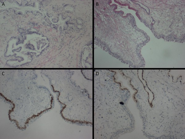
Endocervicosis in the urinary bladder is a rare benign condition. We present a case in a 37-year-old woman with classical clinical and pathological features of endocervicosis. The unusual observation of endocervical-like mucinous epithelium in continuity with the urothelium in addition to fully developed endocervicosis prompted immunohistochemical profiling of the case using antibodies to cytokeratins (AE1/AE3, CK19, CK7, CK5/6, CK20), HBME-1, estrogen receptor (ER) and progesterone receptor (PR) to assess the relationship of the surface mucinous and endocervicosis glandular epithelia. The surface mucinous epithelium, urothelium and endocervicosis glands were immunopositive for AE1/AE3, CK7 and CK19 while CK20 was only expressed by few urothelial umbrella cells. The surface mucinous epithelium was CK5/6 and HBME-1 immunonegative but showed presence of ER and PR. This was in contrast to the urothelium's expression of CK5/6 but not ER and PR. In comparison, endocervicosis glands expressed HBME-1, unlike the surface mucinous epithelium. The endocervicosis epithelium also demonstrated the expected presence of ER and PR and CK5/6 immunonegativity. The slightly differing immunohistochemical phenotypes of the surface mucinous and morphologically similar endocervicosis glandular epithelium is interesting and requires further clarification to its actual nature. The patient has remained well and without evidence of disease 18-months following transurethral resection of the lesion.
Figures
References
-
- Hao H, Tsujimoto M, Tsubamoto H, Komori S, Hirota S. Immunohistochemical phenotype of the urinary bladder endocervicosis: comparison with normal endocervix and well-differentiated mucinous adenocarcinoma of uterine cervix. Pathol Int. 2010;60:528–532. doi: 10.1111/j.1440-1827.2010.02555.x. - DOI - PubMed
MeSH terms
Substances
LinkOut - more resources
Full Text Sources
Medical
Research Materials
Miscellaneous
