Review
. 2012 May;235(1):43-52.
doi: 10.1016/j.expneurol.2011.06.006. Epub 2011 Jun 15.

Myelin associated inhibitors: a link between injury-induced and experience-dependent plasticity

Affiliations
Review

Myelin associated inhibitors: a link between injury-induced and experience-dependent plasticity

Feras Akbik et al. Exp Neurol. 2012 May.

Abstract

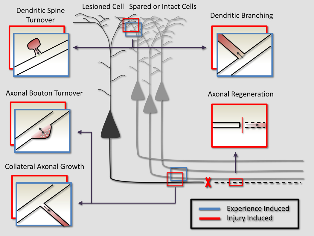
In the adult, both neurologic recovery and anatomical growth after a CNS injury are limited. Two classes of growth inhibitors, myelin associated inhibitors (MAIs) and extracellular matrix associated inhibitors, limit both functional recovery and anatomical rearrangements in animal models of spinal cord injury. Here we focus on how MAIs limit a wide spectrum of growth that includes regeneration, sprouting, and plasticity in both the intact and lesioned CNS. Three classic myelin associated inhibitors, Nogo-A, MAG, and OMgp, signal through their common receptors, Nogo-66 Receptor-1 (NgR1) and Paired-Immunoglobulin-like-Receptor-B (PirB), to regulate cytoskeletal dynamics and inhibit growth. Initially described as inhibitors of axonal regeneration, subsequent work has demonstrated that MAIs also limit activity and experience-dependent plasticity in the intact, adult CNS. MAIs therefore represent a point of convergence for plasticity that limits anatomical rearrangements regardless of the inciting stimulus, blurring the distinction between injury studies and more "basic" plasticity studies.


Figures

Figure 1
Figure 1. Growth Inhibition in the Central Nervous System
There are two classes of known growth inhibitors in the CNS: inhibitors associated with the ECM and those associated with myelin. CSPGs (in brown) are associated with the ECM and astrocytes (in green), and signal at least in part through PTPσ to inhibit growth. Nogo-A, MAG, and OMgp are inhibitors present on the membranes of oligodendrocytes (in red). Nogo-A has two distinct inhibitory domains, a hydrophilic 66 amino acid domain (Nogo-66) and a larger N-terminal domain (amino-Nogo), and immunolocalization studies have demonstrated multiple topologies for Nogo-A that include the one depicted here as well as other topologies, such as extracellular amino-Nogo and intracellular Nogo-66. Despite their lack of sequence homology, all three ligands can bind to two different neuronal (in yellow) receptors, NgR1 and PirB. NgR1 is a GPI linked protein that requires coreceptors to signal intracellularly. It can form one of at least two complexes after binding Nogo-66; NgR1-LINGO-p75 or NgR1-LINGO-TROY. p75 and TROY mediate Nogo-NgR1 dependent activation of the Rho, ROCK signaling cascade that ultimately leads to cytoskeletal changes and growth inhibition. The intracellular signaling cascade between PirB or PTPσ and axonal growth inhibition are not well defined, although both ultimately lead to cytoskeletal changes that mediate growth inhibition. Importantly, both Nogo-A and NgR1 are localized pre and post-synaptically, suggesting an additional role in regulating synaptic plasticity (lower right corner). (CSPGs, chondroitin sulfate proteoglycans; ECM, extracellular matrix; GPI, glycosylphosphatidylinositol; MAG, myelin associated glycoprotein; NgR1, Nogo-Receptor-1; OMgp, oligodendrocyte myelin glycoprotein; PirB, Paired-Immunoglobulin-like Receptor; PTPσ, Protein-Tyrosine-Phosphatase-σ; TROY, TNF-alpha orphan receptor; see text for details.)
Figure 2
Figure 2. Myelin Associated Inhibitors Limit Injury-Induced Growth
(A–E). Anatomical Rearrangements of Lesioned Fibers in Response to Injury are Limited by MAIs. 2–4 month year old female wild-type or mutant mice were subjected to a dorsal hemisection at T6. 4 weeks later, biotin dextran amine (BDA) was unilaterally injected into primary motor cortex to trace the CST, and mice perfused with 4% paraformaldehyde, and 30 µm parasagittal sections were cut on a vibratome. (A) A sagittal section of thoracic spinal cord shows a tight bundle of CST fibers rostral (left) descending until the lesion, and the absence of any fibers caudal to the lesion. Scale bar represents 1 mm. (B) Inset of lesion depicted in (A), demonstrating higher magnification view of abrupt CST interruption at the lesion. Scale bar represents 100 µm. (C) A sagittal section of thoracic spinal cord shows 6 weeks after a dorsal hemisection in mice null for Nogo-A, OMgp, and MAG. Rostral to the lesion, the CST is no longer a tight bundle, reflecting extensive collateral growth of the lesioned tract in response to the injury (arrowhead). Caudal to the lesion, CST fibers can be detected for several millimeters, demonstrating axonal regeneration in an adult mammalian CNS. Scale bar represents 1 mm. (D,E) Insets of spinal cord caudal to the lesion demonstrating regenerating axons fibers (arrowhead). Scale bar represents 100 µm. (F–H). Anatomical Rearrangements of Unlesioned Fibers in Response to Injury are Limited by MAIs. A unilateral lesion to the descending CST at the level of the medullary pyramids (pyramidotomy) was made in either wild-type or NgR1-null adult mice. The intact CST was traced 4 weeks later using unilateral, cortical injections of BDA. Mice were perfused 2 weeks later with 4% paraformaldehyde and 30 µm axial sections were prepared from the cervical spinal cord, caudal to the lesion. Scale bar represents 500 µm. (F,G) In wild-type mouse subjected to a sham surgery (F) or pyramidotomy (G), CST terminals are noted in the ipsilateral grey matter, but very few are seen contralaterally. (H) In NgR1-null mice subject to a pyramidotomy, the CST makes both ipsilateral and contralateral terminals in the grey matter, demonstrating the potential for collateral growth from intact fiber tracts in response to CNS injury. (I–L). Anatomical Rearrangements in Synaptic Structures. Apical tufts of L5 pyramidal cells in S1 of P180 thy-EGFP-M mice were imaged every four days for 28 days through a cranial window. Days 0, 4, 8 and 28 are shown above. Scale bar represents 2 µm. Although most spines persist, a fraction of spines turnover (green asterisk). Of newly formed spines (yellow and red asterisk), only a small percentage persist (yellow asterisk) and make new synapses. Based on their role in limiting a wide spectrum of injury-induced growth, MAIs can potentially regulate dendritic spine elimination, formation, and or persistence.
Figure 3
Figure 3. Myelin Inhibits a Spectrum of Growth in both the Intact and Injured CNS
An injury (red boxes) that severs an axon of a projection neuron leads to a wide spectrum of compensatory growth (represented as a red, emerging process off the original neurite). Anatomical rearrangements in either injured (black) or spared (grey) neurons contribute to functional recovery. This entire spectrum, in both injured and uninjured neurons, is limited by MAIs. In the absence of MAIs, the severed axon could regenerate through the lesion. More rostrally, the injured neuron could sprout collateral dendritic or axonal processes to create multisynaptic connections to bypass the lesion. More subtle changes in the injured neurons synaptic connections, such as differential dendritic spine and axonal bouton turnover and formation, could also redirect signals from the injured neuron around the lesion. In the setting of an injury, spared neurons (grey) can also sprout collateral dendrites or axons, or change their pre or post-synaptic connectivity in an attempt to reroute signaling into the denervated tissue. Experience (blue boxes) stimulates anatomical rearrangements that encode the plastic response to experience. MAIs are known to limit experience-dependent plasticity, and we propose that this is because they also limit anatomical rearrangements in the intact CNS. Experience can be encoded through anatomy by promoting dendritic or axonal collateral growth, or more subtly, the differential stabilization of pre or post-synaptic structures. Ultimately, a common molecular system that regulates cytoskeletal rearrangements links the entire of spectrum of growth, regardless of the initial stimulus (injury or experience).

References

    1. Burns SP, et al. Recovery of ambulation in motor-incomplete tetraplegia. Archives of Physical Medicine and Rehabilitation. 1997;78(11):1169–1172. - PubMed
    1. Geisler FHC, William P, Giacinto Grieco, Devinder Poonian the Sygen Study Group. Measurements and Recovery Patterns in a Multicenter Study of Acute Spinal Cord Injury. Spine. 2001;26(24S):S68–S86. - PubMed
    1. Cajal SRy. Cajal's Degeneration and Regeneration of the Nervous System. Oxford Univeristy Press; 1991. p. 976.
    1. David S, Aguayo A. Axonal elongation into peripheral nervous system "bridges" after central nervous system injury in adult rats. Science. 1981;214(4523):931–933. - PubMed
    1. Richardson PM, McGuinness UM, Aguayo AJ. Axons from CNS neurones regenerate into PNS grafts. Nature. 1980;284(5753):264–265. - PubMed

Publication types

MeSH terms