Review
. 2011 Aug;17(8):1409-21.
doi: 10.1261/rna.2733411. Epub 2011 Jun 23.

Structural aspects of translation termination on the ribosome

Affiliations
Review

Structural aspects of translation termination on the ribosome

Andrei A Korostelev. RNA. 2011 Aug.

Abstract

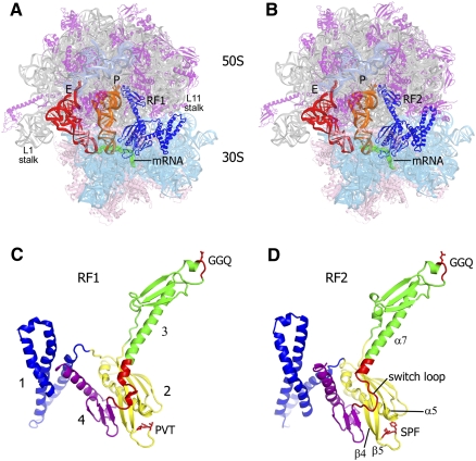
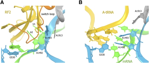
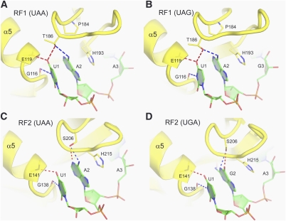
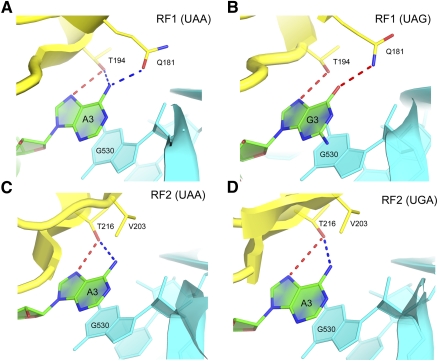
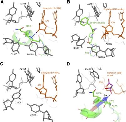
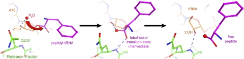
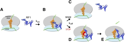
Translation of genetic information encoded in messenger RNAs into polypeptide sequences is carried out by ribosomes in all organisms. When a full protein is synthesized, a stop codon positioned in the ribosomal A site signals termination of translation and protein release. Translation termination depends on class I release factors. Recently, atomic-resolution crystal structures were determined for bacterial 70S ribosome termination complexes bound with release factors RF1 or RF2. In combination with recent biochemical studies, the structures resolve long-standing questions about translation termination. They bring insights into the mechanisms of recognition of all three stop codons, peptidyl-tRNA hydrolysis, and coordination of stop-codon recognition with peptidyl-tRNA hydrolysis. In this review, the structural aspects of these mechanisms are discussed.


Figures

FIGURE 1.
FIGURE 1.
Crystal structures of the 70S translation termination complexes bound with RF1 and RF2. (A,B) 3.2-Å and 3.0-Å structures of the 70S termination complexes bound with RF1 and RF2 (blue) in response to the UAA stop codon (mRNA is shown in green) and in the presence of deacylated P- (orange) and E-site (red) tRNAs. 23S rRNA is shown in gray, 5S rRNA in teal, 50S subunit proteins in magenta, 16S rRNA in cyan, and 30S subunit proteins in pink. (C,D) The structures of RF1 and RF2 in their ribosome-bound conformation, rotated ∼180° from A and B; the structures are colored according to their four-domain organization; GGQ, PVT, and SPF motifs and the switch loop are shown in red.
FIGURE 2.
FIGURE 2.
Differences in the conformations of the decoding center between 70S ribosome complexes bound with (A) RF2 (Korostelev et al. 2008) and (B) a tRNA cognate to the A-site codon (Selmer et al. 2006). RF2 and tRNA are shown in yellow, the switch loop of a release factor is shown in orange, 23S rRNA nucleotide A1913 is shown in gray, 16S rRNA nucleotides G530, A1492, and A1493 are shown in cyan, and mRNA is shown in green.
FIGURE 3.
FIGURE 3.
Interactions of the first two stop-codon nucleotides with release factors RF1 and RF2. (A) Recognition of U1 and A2 of the UAA codon by RF1 (Laurberg et al. 2008) and (B) the UAG codon by RF1 (Korostelev et al. 2010). (C) Recognition of U1 and A2 of the UAA codon by RF2 (Korostelev et al. 2008). (D) Recognition of U1 and G2 of the UGA codon by RF2 (Weixlbaumer et al. 2008). Release factors are shown in yellow, and mRNA is shown in green. (Adapted, with modifications, from Korostelev et al. 2010.)
FIGURE 4.
FIGURE 4.
Interactions of the third stop-codon nucleotide in the 70S translation termination complexes bound with RF1 and RF2. Recognition of the third nucleotide takes place in a G530 pocket of the decoding center. (A,B) Rotation of the conserved Gln181 side chain of RF1 allows it to form hydrogen bonds with either A3 or G3 of the UAA or UAG codons, respectively (Laurberg et al. 2008; Korostelev et al. 2010). (C,D) In RF2, Gln181 is replaced by Val203, restricting the recognition specificity of RF2 to A3. Release factors are shown in yellow, mRNA is shown in green, and 16S rRNA in cyan. (Adapted, with modifications, from Korostelev et al. 2010.)
FIGURE 5.
FIGURE 5.
Conformation of the peptidyl-transferase center depends on the occupancy of the A site. The conformations of the PTC are similar in ribosome complexes, in which the A site is occupied by (A) a release factor (Korostelev et al. 2008) and (B) aminoacyl-tRNA (Voorhees et al. 2009). (C) C2506 and U2585 are found in a different conformation when the 50S subunit A-site is vacant (Selmer et al. 2006). (D) Superposition of a peptidyl-transferase transition-state analog complexed with the 50S subunit (Schmeing et al. 2005a) on the structure of the 70S termination complex (Laurberg et al. 2008). The main-chain amide of Gln230 is positioned to H-bond with the oxyanion of the transition state. The dipole moment of the a-helix likely increases the partial positive charge on the Gln230 backbone NH group and the ability of the NH group to stabilize the negatively charged transition-state intermediate. The dipole moment vector was calculated at Protein Dipole Moments Server (Felder et al. 2007).
FIGURE 6.
FIGURE 6.
Scheme for the mechanism of the peptidyl-tRNA hydrolysis reaction. (Left panel) Nucleophilic attack. The nucleophilic water molecule is positioned for nucleophilic attack at H-bonding distance of the 2′-OH of ribose 76 of the peptidyl-tRNA (Jin et al. 2010). (Center panel) Transition-state stabilization. The oxyanion of the developing tetrahedral transition state is stabilized by the hydrogen bond with the backbone amide NH group of Gln230. (Right panel) Product stabilization. Following hydrolysis, the 3′-hydroxyl leaving group of the deacylated P-site tRNA H-bonds with the backbone amide NH group of Gln230. Crystal structures of RF2-bound 70S complexes in the presence of a peptidyl-tRNA analog (Jin et al. 2010) and deacylated tRNA (Korostelev et al. 2008) were used in the left and right panels, respectively. The transition state (center panel) was modeled by superimposing the 23S rRNA structure of the RF2 termination complex (Korostelev et al. 2008) with the structure of a 50S subunit containing a transition-state analog (Schmeing et al. 2005a). (Adapted, with modifications, from Korostelev et al. 2010.)
FIGURE 7.
FIGURE 7.
Proposed mechanism for coordination of peptidyl-tRNA hydrolysis with stop-codon recognition via a conformational switch in class I release factors. (A,B) Initially, the release factor binds to the ribosome in a catalytically inactive conformation. (C) If a sense codon is located in the A site, the release factor quickly dissociates (Hetrick et al. 2009). (D) If the release factor recognizes a stop codon in the A site, its switch loop along with domain 3 and the decoding center rearrange. Interaction between the switch loop and the switch-loop binding pocket in the decoding center results in tight binding of the release factor to the ribosome. (E) In this catalytically competent conformation, the GGQ motif is inserted in the peptidyl-transferase center and is capable of contributing to catalysis of peptidyl-tRNA ester bond hydrolysis.

References

    1. Alkalaeva EZ, Pisarev AV, Frolova LY, Kisselev LL, Pestova TV 2006. In vitro reconstitution of eukaryotic translation reveals cooperativity between release factors eRF1 and eRF3. Cell 125: 1125–1136 - PubMed
    1. Arkov AL, Murgola EJ 1999. Ribosomal RNAs in translation termination: Facts and hypotheses. Biochemistry (Mosc) 64: 1354–1359 - PubMed
    1. Baierlein C, Krebber H 2010. Translation termination: New factors and insights. RNA Biol 7: 548–550 - PubMed
    1. Ban N, Nissen P, Hansen J, Moore PB, Steitz TA 2000. The complete atomic structure of the large ribosomal subunit at 2.4 A resolution. Science 289: 905–920 - PubMed
    1. Barthelme D, Dinkelaker S, Albers SV, Londei P, Ermler U, Tampe R 2011. Ribosome recycling depends on a mechanistic link between the FeS cluster domain and a conformational switch of the twin-ATPase ABCE1. Proc Natl Acad Sci 108: 3228–3233 - PMC - PubMed

LinkOut - more resources