. 2011 Sep 29;118(13):3708-14.
doi: 10.1182/blood-2011-01-332676. Epub 2011 Jun 23.

Histones induce rapid and profound thrombocytopenia in mice

Affiliations

Histones induce rapid and profound thrombocytopenia in mice

Tobias A Fuchs et al. Blood. .

Abstract

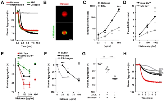
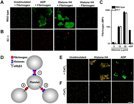
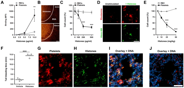
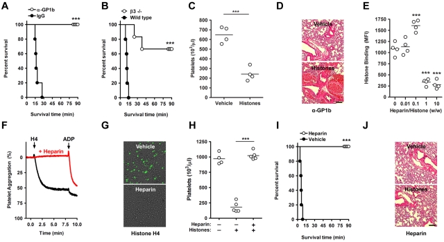
Histones are released from dying cells and contribute to antimicrobial defense during infection. However, extracellular histones are a double-edged sword because they also damage host tissue and may cause death. We studied the interactions of histones with platelets. Histones bound to platelets, induced calcium influx, and recruited plasma adhesion proteins such as fibrinogen to induce platelet aggregation. Hereby fibrinogen cross-linked histone-bearing platelets and triggered microaggregation. Fibrinogen interactions with αIIbβ3 integrins were not required for this process but were necessary for the formation of large platelet aggregates. Infused histones associated with platelets in vivo and caused a profound thrombocytopenia within minutes after administration. Mice lacking platelets or αIIbβ3 integrins were protected from histone-induced death but not from histone-induced tissue damage. Heparin, at high concentrations, prevented histone interactions with platelets and protected mice from histone-induced thrombocytopenia, tissue damage, and death. Heparin and histones are evolutionary maintained. Histones may combine microbicidal with prothrombotic properties to fight invading microbes and maintain hemostasis after injury. Heparin may provide an innate counter mechanism to neutralize histones and diminish collateral tissue damage.


Figures

Figure 1
Figure 1
Histone-induced platelet aggregation requires β3-integrins. (A) Histones induce platelet aggregation. PRP was mixed at t = 0 minutes with histones (200 μg/mL), ADP (50μM), or collagen (25 μg/mL) or left unstimulated. Histones induced aggregation as efficiently as ADP or collagen. (B) Histones bind to platelets. Fluorescence microscopy of platelets labeled in red incubated with BSA or histones labeled fluorescently in green. Histones localize to the platelet surface. Scale bar = 2 μm. (C) Quantification of histone or BSA binding to washed platelets by flow cytometry (***compared with BSA; n = 3). (D) Histones induce calcium influx into platelets. Platelets loaded with the calcium-sensitive dye Fluo-4 and stimulated with indicated concentrations of histones for 5 minutes. Platelets were resuspended in medium with or without CaCl2, and fluorescence was analyzed by flow cytometry (**compared with or without Ca2+; n = 6). (E) Comparison of histone-induced aggregation of control mice (C57, red), CalDAG-GEF1 (black), or β3-integrin (green) deficient platelets, 5 minutes after stimulation with indicated concentrations of histones or ADP (50μM). CalDAG-GEF1 deficiency leads to an impaired platelet aggregation in response to histones, but it is less severe than β3-integrin deficiency. (F) Histone-induced platelet aggregation requires plasma proteins. Dose-dependent response of platelets to histones in the presence of plasma (black circles), 200 μg/mL fibrinogen (gray circles), or the response of washed platelets with buffer (white circle); n = 3; ***plasma compared with fibrinogen. (G) Exogenous CaCl2 (2mM) enhanced HiPA. Data show extent of platelet aggregation 3 minutes after stimulation (n = 4). (H) Aggregation of washed platelets stimulated with 1μM recombinant histone H1, H2A, H2B, H3, or H4 in the presence of 200μg/mL fibrinogen. Histone H4 induced platelet aggregation potently. Data presented are representative of ≥ 3 independent experiments. *P < .05, **P < .01, and ***P < .001.
Figure 2
Figure 2
Histones recruit fibrinogen and cause platelet aggregation through mechanisms dependent and independent of αIIbβ3 integrins. Fluorescence imaging of wild-type (A) or β3-integrin–deficient (B) platelets stimulated in the presence or absence of fibrinogen with ADP (50μM) or recombinant histone H4 (10 μg/mL). Histone H4 or ADP induce large platelet aggregates of wild-type but not β3-integrin–deficient platelets. Recombinant histone H4 but not ADP induces microaggregation of β3-integrin–deficient platelets. This microaggregation depended on the presence of fibrinogen. (C) Histones induce β3-integrin–independent fibrinogen binding to platelets. Washed platelets stimulated with the indicated concentrations of histones or ADP (50μM) were incubated with fluorescent fibrinogen and analyzed by flow cytometry. (D) Hypothesized interactions of fibrinogen with αIIbβ3 and histones in HiPA. Fibrinogen can bind to αIIbβ3 and histones on platelets. Fluorescence imaging of wild-type (green) or β3-integrin–deficient (red) platelets mixed with fibrinogen in response to ADP or histones in the presence (E) or absence (F) of CaCl2. Recombinant histone H4 but not ADP induced the formation of large platelet aggregates consisting of wild-type and β3-deficient platelets. This process depended on exogenous CaCl2. Scale bar = 100 μm. Data presented are representative of ≥ 3 independent experiments.
Figure 3
Figure 3
Histones induce thrombocytopenia. Histones bind to platelets in blood. (A) Fluorescent histones were mixed at the indicated concentrations with blood. Flow cytometric analysis showed that histones bind preferentially to platelets over RBCs in blood (**compared with RBCs; n = 5). (B) Perfusion of Rhodamine 6G–labeled platelets in blood over a histone-coated surface shows platelet binding to histones. Areas covered with BSA only did not bind platelets. Scale bar = 100 μm. (C) Histones induce thrombocytopenia in blood in vitro. Single platelet and RBC counts of blood mixed with indicated concentrations of histones. At concentrations of 250 μg/mL histones induced thrombocytopenia in vitro (***compared with RBCs; n = 3). (D) Microscopic analysis of Rhodamine 6G–labeled platelets in blood before (Unstimulated) and after stimulation with Alexa 488–labeled histones (+Histones). Histones induce and localize to platelet aggregates in blood. Scale bar = 50 μm. Histones induce thrombocytopenia in vivo. (E) Platelet and RBC counts of mice 10 minutes after infusion with indicated sublethal concentrations of histones (***compared with RBCs; n = 3). Histone infusion caused dose-dependent depletion of platelets from circulation. (F) Determination of tail bleeding time. Mice infused with histones (50 mg/kg) but not vehicle showed prolonged bleeding time. Histones associate with platelets in vivo. Immunostaining of lungs dissected 10 minutes after infusion with histones (G-I; 50 mg/kg) or vehicle (J). Lungs were stained for CD41 (platelets), histone H3 (histones), and DNA. Platelets (G) and histones (H) colocalized in mice infused with histones (I) but not in mice infused with vehicle (J). Under these conditions, histone staining of nuclei was below the detection limit. Scale bar = 50 μm. Data presented are representative of ≥ 3 independent experiments. **P < .01, ***P < .001.
Figure 4
Figure 4
Heparin prevents histone-induced thrombocytopenia, tissue damage, and mortality. Platelets contribute to histone-induced mortality. (A) Platelet depletion protects from histone-induced death. Infusion of platelet-depleting antibodies (anti-GP1b) but not control IgG protected mice from a lethal dose of histones (60 mg/kg). (B) β3-Integrin deficiency partially protects from histone-induced death. Survival time of wild-type and β3-integrin–deficient mice infused with histones (60 mg/kg). (C) β3-Integrin deficiency does not protect from histone-induced thrombocytopenia (25 mg/kg). Platelet counts of β3−/− mice 10 minutes after infusion with vehicle or histones. (D) Platelet depletion does not protect from histone-induced tissue damage. H&E stainings of lungs from platelet-depleted mice (anti-Gp1b) 90 minutes after the infusion of vehicle or histones. Hemorrhage indicates histone-induced damage of the vascular endothelium. Scale bars = 250 μm. (E) At high concentrations, heparin prevents the interaction of histones with platelets. Flow cytometric analysis of histone binding to platelets in the presence of indicated concentrations of heparin (***compared with 0). (F) Heparin prevents HiPA. Aggregometry of platelets mixed with plasma in the presence (red) or absence (black) of heparin and in response to recombinant histone H4 or ADP. (G) Heparin neutralizes histone-induced cytotoxicity. Overlays of SytoxGreen fluorescence and phase-contrast images. A murine endothelial cell line was incubated in the presence or absence of heparin (100 μg/mL) with recombinant histone H4 (20 μg/mL). Cell death was detected by staining with the cell-impermeable DNA dye SytoxGreen. The presence of heparin prevented cytotoxicity. (H) Heparin prevents histone-induced thrombocytopenia. Platelet counts of untreated mice, mice infused with histones (50 mg/kg), and mice treated with heparin (50 mg/kg) before histone infusion. (I) Heparin prevents histone-induced lethality. Survival of mice treated with 50 mg/kg heparin or vehicle before infusion of 75 mg/kg histones. (J) Heparin prevents histone-induced hemorrhage in lungs. H&E stainings of lungs from mice treated with heparin after the infusion of vehicle or histones. Data are representative of ≥ 3 independent experiments. *P < .05, **P < .01, and ***P < .001.

Comment in

Similar articles

Cited by

References

    1. Brinkmann V, Reichard U, Goosmann C, et al. Neutrophil extracellular traps kill bacteria. Science. 2004;303(5663):1532–1535. - PubMed
    1. Hirsch JG. Bactericidal action of histone. J Exp Med. 1958;108(6):925–944. - PMC - PubMed
    1. Xu J, Zhang X, Pelayo R, et al. Extracellular histones are major mediators of death in sepsis. Nat Med. 2009;15(11):1318–1321. - PMC - PubMed
    1. Zeerleder S, Zwart B, Wuillemin WA, et al. Elevated nucleosome levels in systemic inflammation and sepsis. Crit Care Med. 2003;31(7):1947–1951. - PubMed
    1. Fuchs TA, Brill A, Duerschmied D, et al. Extracellular DNA traps promote thrombosis. Proc Natl Acad Sci U S A. 2010;107(36):15880–15885. - PMC - PubMed

Publication types

MeSH terms