. 2011 Sep;301(3):G475-86.
doi: 10.1152/ajpgi.00071.2011. Epub 2011 Jun 23.

Specific effects of BCL10 Serine mutations on phosphorylations in canonical and noncanonical pathways of NF-κB activation following carrageenan

Affiliations

Specific effects of BCL10 Serine mutations on phosphorylations in canonical and noncanonical pathways of NF-κB activation following carrageenan

Sumit Bhattacharyya et al. Am J Physiol Gastrointest Liver Physiol. 2011 Sep.

Abstract

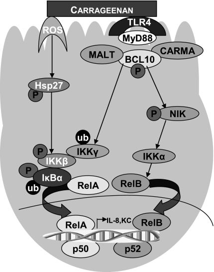
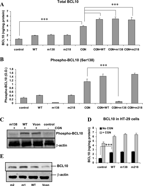
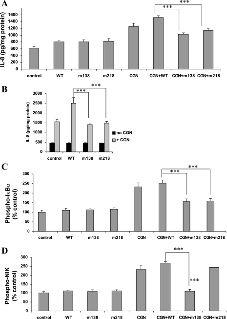
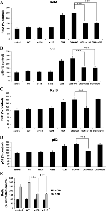
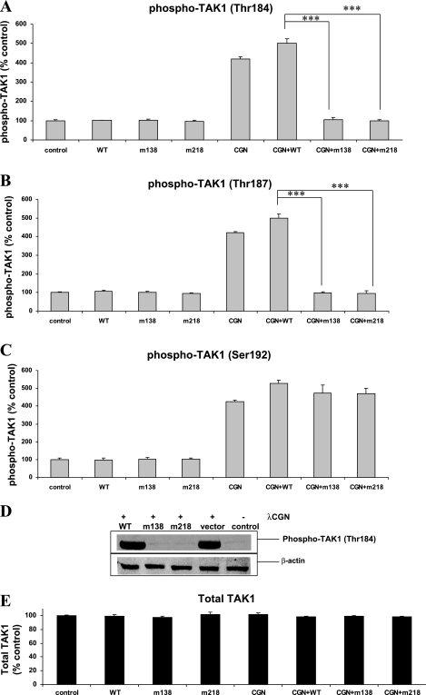
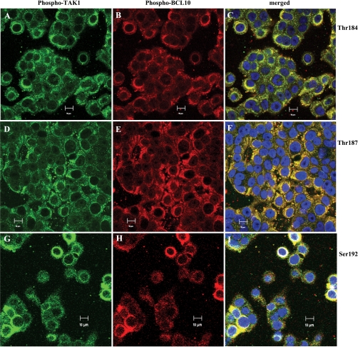
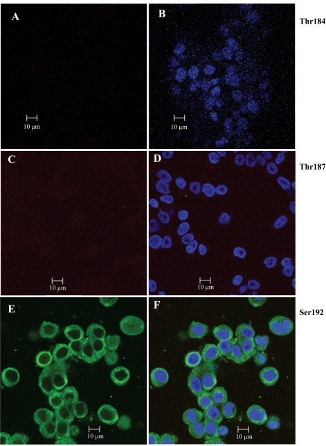
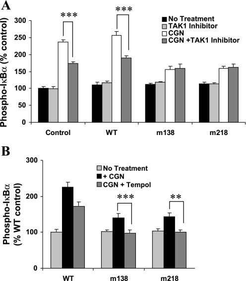
To determine the impact of B cell leukemia/lymphoma (BCL) 10 on the phosphorylation of crucial mediators in NF-κB-mediated inflammatory pathways, human colonic epithelial cells were exposed to carrageenan (CGN), a sulfated polysaccharide commonly used as a food additive and known to induce NF-κB nuclear translocation by both canonical and noncanonical pathways. Phosphorylations of intermediates in inflammatory cascades, including NF-κB-inducing kinase (NIK) at Thr(559), transforming growth factor-β-activating kinase (TAK) 1 at Thr(184), Thr(187), and Ser(192), and inhibitory factor κBα (IκBα) at Ser(32), were examined following mutation of BCL10 at Ser(138) and at Ser(218). Specific phosphoantibodies were used for detection by enzyme-linked immunosorbent assay, immunoblot, and confocal microscopy of differences in phosphorylation following transfection by mutated BCL10. Both mutations demonstrated dominant-negative effects, with inhibition of phospho(Ser(32))-IκBα to less than control levels. Both of the BCL10 mutations reduced the CGN-induced increases in nuclear RelA and p50, but only the Ser(138) mutation inhibited the CGN-induced increases in nuclear RelB and p52 and in NIK Thr(559) phosphorylation. Hence, the phosphorylation of BCL10 Ser(138), but not Ser(218), emerged as a critical event in activation of the noncanonical pathway of NF-κB activation. Either BCL10 Ser(138) or Ser(218) mutation inhibited the phosphorylation of TAK1 at Thr(184) and at Thr(187), but not at Ser(192). These findings indicate that BCL10 phosphorylations act upstream of phosphorylations of NIK, TAK1, and IκBα and differentially affect the canonical and noncanonical pathways of NF-κB activation.


Figures

Fig. 1.
Fig. 1.
Schematic of the inflammatory cascades activated by exposure to carrageenan (CGN) in NCM460 cells. Three distinct pathways of CGN-induced inflammation in colonic epithelial cells include 1) a Toll-like receptor (TLR) 4-B cell leukemia/lymphoma (BCL) 10 pathway that leads to nuclear TLR4-BCL10-inhibitory factor κB kinase (IKK) γ-IKKβ-inhibitory factor κBα (IκBα)-NF-κB (RelA; p65); 2) a TLR4-BCL10 pathway leading to nuclear TLR4-BCL10-NF-κB-inducing kinase (NIK)-IKKα-NF-κB (RelB); and 3) a reactive oxygen species (ROS)-mediated pathway leading to nuclear RelA. Ub, ubiquitin; IL, interleukin; Hsp, heat-shock protein; IRAK, interleukin-1β-activating kinase; KC, mouse IL-8 homolog; CARMA, CARD-containing membrane-associated guanylate kinase; MALT, mucosa-associated lymphoid tissue.
Fig. 2.
Fig. 2.
Effects of BCL10 mutations on total BCL10 and phospho-BCL10. A: CGN treatment (1 μg/ml × 24 h) of NCM460 cells increased BCL10 from 1.26 ± 0.11 to 3.92 ± 0.15 ng/mg protein (P < 0.001, 1-way ANOVA with Tukey-Kramer posttest) when quantified by enzyme-linked immunosorbent assay (ELISA). After transfection with either wild-type (WT) BCL10 or BCL10 with S138G or S218G mutations, CGN increases to ∼4.2 times the baseline level of total BCL10. All increases in total BCL10 following CGN were statistically significant (P < 0.001). Following transfection with either WT or the mutant BCL10s, the CGN-induced increases were significantly higher than following CGN stimulation of the control cells (P < 0.001). (Unless stated otherwise, statistical significance was determined by 1-way ANOVA with Tukey-Kramer posttest for multiple comparisons.) B: increases in phospho-BCL10 were measured using a phospho-BCL10 (Ser138) antibody and cell-based ELISA assay. CGN (1 μg/ml × 24 h) caused significant increases, except in the cells transfected with the S138G mutation, demonstrating the effectiveness of the transfection in reducing phospho(Ser138)-BCL10. The S138G BCL10 mutant appears to act as a dominant negative and suppresses the phosphorylation of the endogenous BCL10. C: representative Western blot demonstrates the increase in phospho-BCL10 (Ser138) that followed transfection with WT BCL10 and exposure to CGN in the NCM460 cells. Following transfection with the S138G mutant, no increase in phospho-BCL10 occurred. m138, S138G BCL10 mutant; m218, S218G BCL10 mutant. D: when HT-29 cells were transfected with BCL10 WT, S138G, and S218G mutations, BCL10 increased significantly (P < 0.001), suggesting greater transfection efficiency in the HT-29 cells than in the NCM460 cells. CGN exposure produced further increases in total BCL10 in the transfected cells (P < 0.001). E: in HT-29 cells, immunoblot also demonstrated increases in BCL10 following transfection with the WT and mutant BCL10 constructs. m1, m138=S138G BCL10 mutant; m2, m218=S218G BCL10 mutant; Vcon, empty vector control. *** P ≤ 0.001.
Fig. 3.
Fig. 3.
Effects of BCL10 mutations on IL-8, phospho-IκBα, and phospho-NIK. A: following CGN exposure, IL-8 secretion in the NCM460 cells transfected with WT BCL10 increased from 808 ± 28 to 1,520 ± 54 pg/mg of protein (P < 0.001). Following transfection with the BCL10 mutants, the increases in IL-8 were significantly reduced (P < 0.001). B: in the HT-29 cells, IL-8 secretion increased following transfection with WT BCL10 and exposure to CGN, but the increases were reduced significantly following transfection with the mutant BCL10 constructs (P < 0.001). C: in the NCM460 cells, the CGN-induced increases in phospho-IκBα (Ser32) were reduced to ∼1.6 times the baseline level by both of the BCL10 mutations from ∼2.5 times baseline in the CGN-treated cells transfected by WT BCL10 (P < 0.001). D: following transfection with BCL10 with the S138G mutation in the NCM460 cells, the CGN-induced increase in phospho(Thr559)-NIK was inhibited (P < 0.001), but the S218G mutation did not inhibit the CGN-induced increase in phospho-NIK. These findings are consistent with a vital role for BCL10 Ser138 phosphorylation in the noncanonical pathway of NF-κB activation. ***P ≤ 0.001.
Fig. 4.
Fig. 4.
BCL10 mutations have differential effects on NF-κB family members. A: NCM460 cells were transfected with either WT or mutated BCL10 and exposed to λ-CGN (1 μg/ml × 24 h), and NF-κB components were measured by an NF-κB oligonucleotide assay. Nuclear RelA increased to 2.52 ± 0.03 times the baseline value following CGN and transfected by WT BCL10, but declined significantly, to ∼1.5 times the baseline value (P < 0.001), following transfection with either of the BCL10 mutants. B: similar to the changes in RelA, p50 increased to 2.67 ± 0.22 times the baseline value when transfected with WT BCL10 and treated with CGN, but to only ∼1.60 ± 0.05 (S138G) and 1.5 6 ± 0.12 (S218G) times the baseline values when transfected with either of the mutants (P < 0.001). C: following transfection with either the WT BCL10 or the S218G mutant, RelB increased to ∼1.5 times the baseline value. When transfected with the S138G BCL10, this increase in RelB was inhibited (P < 0.001), consistent with the requirement for BCL10 Ser138 for activation of the noncanonical pathway of NF-κB. D: corresponding to the changes in RelB, p52 increased to ∼1.65 times the baseline value following transfection with either WT BCL10 or the S218G mutant and CGN exposure. However, following transfection with the S138G mutation, no increase in p52 occurred (P < 0.001), consistent with the requirement of BCL10 Ser138 for the noncanonical pathway and the dominant-negative effect of the mutant BCL10. E: in the HT-29 cells, RelA increased to 2.5 ± 0.3 times the baseline value following exposure to CGN. Following transfection with WT BCL10 and CGN exposure (1 μg/ml × 24 h), RelA increased to 3.0 ± 0.2 times the baseline but declined to 1.6 times baseline when transfected with either of the mutant BCL10 constructs (P < 0.001) ***P ≤ 0.001.
Fig. 5.
Fig. 5.
BCL10 mutations inhibit phosphorylation of transforming growth factor-β-activating kinase (TAK) 1 Thr184 and Thr187 but not Ser192. A–C: CGN treatment (1 μg/ml × 24 h) of NCM460 cells yielded an increase to 4.2 times the baseline level in phospho-TAK1 (Thr184, Thr187, or Ser192) in control, untransfected cells. Following transfection by WT BCL10 and CGN treatment, these increases were 5.0, 5.0, and 5.3 times the baseline level, respectively (P < 0.001). Following transfection by the mutated BCL10 and CGN treatment, no increases in phospho-TAK1 (Thr184 or Thr187) occurred, but the increase in phospho-TAK1 (Ser192) was not inhibited. Following CGN, phospho-TAK1(Ser192) increases ranged from 4.2 to 5.3 times the baseline levels. D: representative Western blot of phospho-TAK1(Thr184) demonstrates the CGN-induced increase in phospho-TAK1(Thr184) in the control and WT transfectant, but not following transfection with the BCL10 mutant constructs. E: total TAK1 was not affected by treatment with CGN or transfection with BCL10 WT or mutated BCL10. ***P ≤ 0.001.
Fig. 6.
Fig. 6.
Confocal images of phospho-BCL10 and phospho(Thr184,Thr187,Ser192)TAK1 demonstrate colocalization following transfection by WT BCL10. A-I: the phospho-TAK1 findings are supported by confocal images acquired using specific phospho-TAK1 and phospho-BCL10 (Ser138) antibodies. In images A-I, NCM460 cells were transfected with the WT BCL10. Specific phospho-TAK1 antibodies were used, showing phosphorylated (p)-Thr184 (A), p-Thr187 (D), and p-Ser192 (G) as green. Phospho-BCL10 (Ser138) antibody was used as described in materials and methods and appears red (B, E, and H). Phospho-BCL10 (Ser138) and phospho-TAK1 colocalized and stained yellow, as seen in C, F, and I. Nuclei are stained blue by 4′,6-diamidino-2-phenylindole (DAPI). Marker = 10 μm.
Fig. 7.
Fig. 7.
Confocal images following transfection with mutated BCL10 (S138G). A–F: NCM460 cells were transfected with mutated BCL10 (S138G), and staining was repeated as above. Phospho-TAK1 (Thr184 and Thr187) are absent in images A–B and C–D, respectively. In contrast, images E and F demonstrate the presence of phospho-TAK1 (Ser192) (green). Distinct phospho-BCL10 (Ser138) immunostaining (red) is absent, and merged images show no yellow immunostaining. Nuclei stain blue using DAPI. Marker = 10 μm.
Fig. 8.
Fig. 8.
Effect of TAK1 inhibitor and ROS inhibitor on phospho-IκBα following CGN and BCL10 mutation. A: TAK1 inhibition significantly reduced the CGN-induced increases in phospho-IκBα in the control and following transfection with WT BCL10 (P < 0.001). Following transfection with the mutated BCL10 constructs, the TAK1 inhibitor had no effect on the CGN-induced increase in phospho-IκBα, indicating a requirement for BCL10 Ser138 or Ser218 for the effect of TAK1 on CGN-induced phosphorylation of IκBα. B: when NCM460 cells were transfected with the mutant BCL10 and exposed to the ROS quencher 2,2,6,6-tetramethylpiperidinyloxy (Tempol) before CGN treatment, the CGN-induced increase in phospho-IκBα was completely inhibited, in contrast to the lack of effect of the TAK1 inhibitor. The effect of Tempol is consistent with involvement of both a BCL10-mediated pathway and an ROS-mediated pathway on the CGN-induced increase in phospho-IκBα. **P ≤ 0.01 and ***P ≤ 0.001.

Similar articles

Cited by

References

    1. Bhattacharyya S, Borthakur A, Dudeja PK, Tobacman JK. Lipopolysaccharide-induced activation of NF-kappaB non-canonical pathway requires BCL10 serine 138 and NIK phosphorylations. Exp Cell Res 316: 3317–3327, 2010 - PMC - PubMed
    1. Bhattacharyya S, Borthakur A, Pant N, Dudeja PK, Tobacman JK. Bcl10 mediates LPS-induced activation of NF-kappaB and IL-8 in human intestinal epithelial cells. Am J Physiol Gastrointest Liver Physiol 293: G429–G437, 2007 - PubMed
    1. Bhattacharyya S, Borthakur A, Tyagi S, Gill R, Chen ML, Dudeja PK, Tobacman JK. B-cell CLL/lymphoma 10 (BCL10) is required for NF-kappaB production by both canonical and noncanonical pathways and for NF-kappaB-inducing kinase (NIK) phosphorylation. J Biol Chem 285: 522–530, 2010 - PMC - PubMed
    1. Bhattacharyya S, Dudeja PK, Tobacman JK. Carrageenan-induced NFkappaB activation depends on distinct pathways mediated by reactive oxygen species and Hsp27 or by Bcl10. Biochim Biophys Acta 1780: 973–982, 2008 - PMC - PubMed
    1. Bhattacharyya S, Dudeja PK, Tobacman JK. Lipopolysaccharide activates NF-kappaB by TLR4-Bcl10-dependent and independent pathways in colonic epithelial cells. Am J Physiol Gastrointest Liver Physiol 295: G784–G790, 2008 - PubMed

Publication types

MeSH terms