. 2011 Jul;121(7):2845-54.
doi: 10.1172/JCI57324. Epub 2011 Jun 23.

Intrarenal dopamine deficiency leads to hypertension and decreased longevity in mice

Affiliations

Intrarenal dopamine deficiency leads to hypertension and decreased longevity in mice

Ming-Zhi Zhang et al. J Clin Invest. 2011 Jul.

Abstract

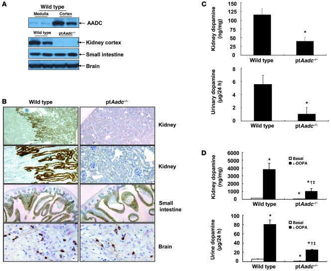
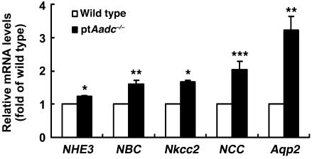
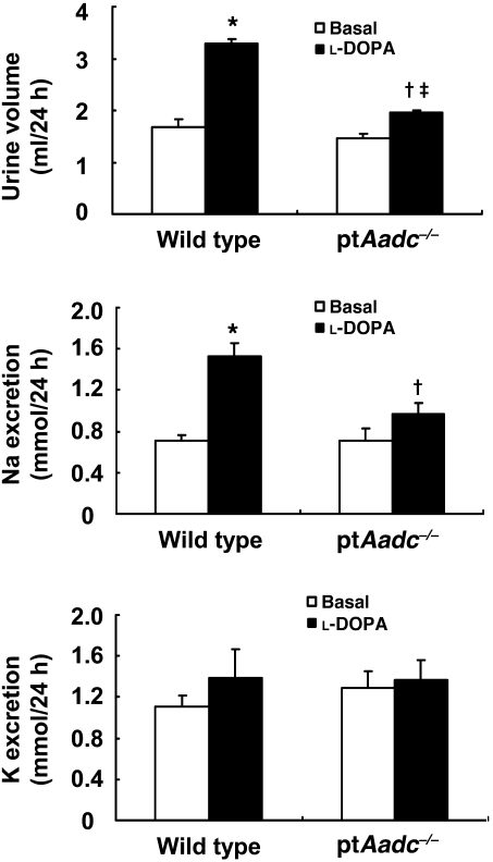
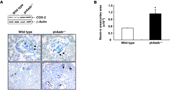
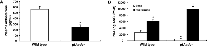
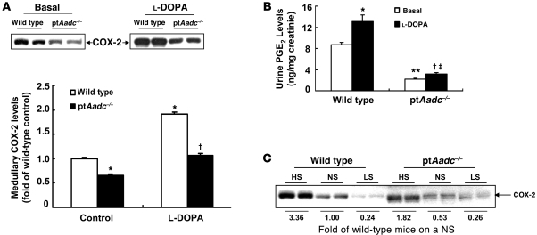
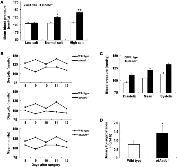
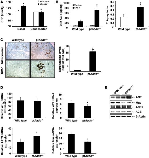
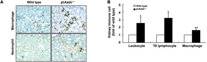
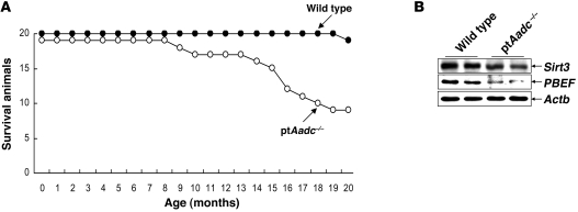
In addition to its role as an essential neurotransmitter, dopamine serves important physiologic functions in organs such as the kidney. Although the kidney synthesizes dopamine through the actions of aromatic amino acid decarboxylase (AADC) in the proximal tubule, previous studies have not discriminated between the roles of extrarenal and intrarenal dopamine in the overall regulation of renal function. To address this issue, we generated mice with selective deletion of AADC in the kidney proximal tubules (referred to herein as ptAadc-/- mice), which led to selective decreases in kidney and urinary dopamine. The ptAadc-/- mice exhibited increased expression of nephron sodium transporters, decreased natriuresis and diuresis in response to l-dihydroxyphenylalanine, and decreased medullary COX-2 expression and urinary prostaglandin E2 excretion and developed salt-sensitive hypertension. They had increased renin expression and altered renal Ang II receptor (AT) expression, with increased AT1b and decreased AT2 and Mas expression, associated with increased renal injury in response to Ang II. They also exhibited a substantially shorter life span compared with that of wild-type mice. These results demonstrate the importance of the intrarenal dopaminergic system in salt and water homeostasis and blood pressure control. Decreasing intrarenal dopamine subjects the kidney to unbuffered responses to Ang II and results in the development of hypertension and a dramatic decrease in longevity.


Figures

Figure 1
Figure 1. AADC was selectively deleted in the renal proximal tubule in ptAadc–/– mice.
(A) AADC was primarily expressed in kidney cortex in wild-type mice. AADC expression was reduced in kidney cortex but not in small intestine or brain in ptAadc–/– mice. (B) Representative photomicrographs indicate reduced AADC expression in renal proximal tubule epithelia but not in small intestine or brain in ptAadc–/– mice. Original magnification: ×25 (first row); ×100 (second row); ×63 (third row); ×250 (fourth row). (C) Kidney dopamine levels and urinary dopamine excretion were significantly decreased in ptAadc–/– mice (*P < 0.01; n = 5 in each group). (D) Kidney dopamine levels and urinary dopamine excretion in response to l-DOPA were significantly attenuated in ptAadc–/– mice (*P < 0.01 vs. basal wild type, P < 0.01 vs. basal ptAadc–/–, P < 0.01 vs. l-DOPA wild type; n = 4 in each group).
Figure 2
Figure 2. Renal mRNA levels of sodium transporters were increased in ptAadc–/–mice.
Four-week-old wild-type and ptAadc–/– mice were studied (*P < 0.001, **P < 0.01, ***P < 0.02; n = 4 in each group).
Figure 3
Figure 3. l-DOPA–induced diuresis and natriuresis were attenuated in ptAadc–/– mice.
Three-month-old male mice were studied. l-DOPA induced significantly less increased urine volume and sodium excretion in ptAadc–/– mice than in wild-type mice (urine volume, *P < 0.001 vs. basal wild type, P < 0.01 vs. basal ptAadc–/–, P < 0.001 vs. l-DOPA–treated wild type) (sodium excretion, *P < 0.005 vs. basal wild type, P < 0.05 vs. l-DOPA–treated wild type) (n = 6 in each group).
Figure 4
Figure 4. Renal cortical COX-2 and renin expression increased in ptAadc–/– mice.
(A) Immunoblotting showed increased renal cortical COX-2 expression in ptAadc–/– mice. Representative photomicrographs indicated increased renal cortical COX-2 and renin expression in ptAadc–/–mice. Original magnification: ×400 (COX-2); ×100 (renin). (B) Quantitative image analysis indicated increased renal (ir) renin expression in ptAadc–/– mice (*P < 0.01; n = 4).
Figure 5
Figure 5. Plasma aldosterone levels and renin activity were suppressed in ptAadc–/– mice.
(A) Plasma aldosterone levels were significantly lower in ptAadc–/– than in wild-type mice (*P < 0.001; n = 6). (B) PRA was lower in untreated ptAadc–/– mice than in wild-type mice but increased more in ptAadc–/– mice than in wild-type mice after hydralazine stimulation (*P < 0.01 vs. basal wild type, P < 0.001 vs. basal ptAadc–/–, P < 0.001 vs. hydralazine stimulated wild type; n = 6 in each group).
Figure 6
Figure 6. Renal medullary COX-2 was inhibited in ptAadc–/– mice.
(A) Renal medullary COX-2 levels were lower in ptAadc–/– mice than wild-type mice, with or without l-DOPA stimulation (*P < 0.01 vs. basal wild type, P < 0.001 vs. l-DOPA treated wild type; n = 4 in each group). (B) Urinary PGE2 levels were significantly lower in ptAadc–/– mice than wild-type mice, with or without l-DOPA stimulation (*P < 0.05 vs. basal wild type, **P < 0.01 vs. basal wild type, P < 0.05 vs. basal ptAadc–/–, P < 0.005 vs. l-DOPA–treated wild type; n = 4 in each group). (C) High-salt diet–induced (HS-induced) renal medullary COX-2 elevations were attenuated in ptAadc–/– mice. Also shown is densitometric quantification of COX-2 immunoreactive protein in response to alterations of dietary salt intake represented as fold of expression of wild-type mice on a normal-salt diet (NS). LS, low-salt diet.
Figure 7
Figure 7. ptAadc–/– mice developed salt-sensitive hypertension.
(A) MBP was similar between 3-month-old ptAadc–/– mice and wild-type mice on a low-salt diet. Increasing dietary salt intake had no effect on MBP in wild-type mice but led to progressive increases in MBP in ptAadc–/– mice (*P < 0.01 vs. wild type on a normal-salt diet, P < 0.05 vs. wild type on a high-salt diet, P < 0.005 vs. ptAadc–/– on a normal-salt diet; n = 9 in each group). (B) Dynamics of systolic, diastolic, and MBP measured by radiotelemetry from 8 to 12 days after surgery (n = 4). (C) MBPs measured by radiotelemetry from 8 to 12 days after surgery. (D) Male wild-type and ptAadc–/– mice were fed an 8% high-salt diet for 4 weeks, and 24-hour urine was collected for measurement of F2-isoprostanes (*P < 0.05; n = 6).
Figure 8
Figure 8. The intrarenal renin-angiotensin system was altered in ptAadc–/– mice.
(A) Antagonism of AT1 receptors with candesartan reduced SBP in ptAadc–/– mice to levels seen in wild-type mice (*P < 0.01 vs. basal wild type, P < 0.01 vs. basal ptAadc–/–; n = 6 in each group). (B) Ang II infusion (0.9 mg/kg/d) for 4 weeks led to more significant increases in albuminuria (*P = 0.012) and tubulointerstitial (TI) injury (P = 0.03) in ptAadc–/– mice than wild-type mice (n = 5). ACR, albumin creatinine ratio. (C) The expression of nitrotyrosine (a marker of oxidative stress) and KIM-1 (a marker of kidney injury) was much higher in Ang II–treated ptAadc–/– mice than Ang II–treated wild-type mice. Original magnification: ×63. Quantitative image analysis indicated nitrotyrosine levels were significantly higher in Ang II–treated ptAadc–/– mice than Ang II–treated wild-type mice (*P < 0.01; n = 4). (D) There were increased mRNA levels of AT1b (*P < 0.00001) but decreased mRNA levels of AT2 (*P < 0.00001) and Mas (P < 0.0001) in ptAadc–/– mice compared with those in wild-type mice (n = 4). (E) Immunoblotting indicated increased angiotensinogen (AGT) but decreased Mas protein levels in ptAadc–/– mice.
Figure 9
Figure 9. Immune cell infiltration was higher in old ptAadc–/– mice than old wild-type mice.
(A) Representative photomicrographs indicated more macrophage and neutrophil infiltration in 20-month-old ptAadc–/– mice than 20-month-old wild-type mice. Original magnification: ×160. (B) FACS analysis indicated increased leukocyte, T8 lymphocyte, and macrophage infiltration in old ptAadc–/– mice than old wild-type mice (*P < 0.05, **P < 0.01; n = 3).
Figure 10
Figure 10. Renal dopamine deficiency was associated with shorter life spans.
(A) At 20 months of age, 10 out of 19 ptAadc–/– mice had died, while only 1 out of 20 wild-type mice had died. (B) Immunoblotting showed reduced levels of prosurvival genes Sirt3 and PBEF in ptAadc–/– mice.

References

    1. Felder RA, Jose PA. Mechanisms of disease: the role of GRK4 in the etiology of essential hypertension and salt sensitivity. Nat Clin Pract Nephrol. 2006;2(11):637–650. doi: 10.1038/ncpneph0301. - DOI - PubMed
    1. Aperia AC. Intrarenal dopamine: a key signal in the interactive regulation of sodium metabolism. Annu Rev Physiol. 2000;62:621–647. doi: 10.1146/annurev.physiol.62.1.621. - DOI - PubMed
    1. Banday AA, Lokhandwala MF. Dopamine receptors and hypertension. Curr Hypertens Rep. 2008;10(4):268–275. doi: 10.1007/s11906-008-0051-9. - DOI - PubMed
    1. Zeng C, Jose PA. Dopamine receptors: important antihypertensive counterbalance against hypertensive factors. Hypertension. 2011;57(1):11–17. doi: 10.1161/HYPERTENSIONAHA.110.157727. - DOI - PMC - PubMed
    1. Quinones H, Collazo R, Moe OW. The dopamine precursor L-dihydroxyphenylalanine is transported by the amino acid transporters rBAT and LAT2 in renal cortex. Am J Physiol Renal Physiol. 2004;287(1):F74–F80. doi: 10.1152/ajprenal.00237.2003. - DOI - PubMed

Publication types

MeSH terms