. 2011 Jul;121(7):2855-62.
doi: 10.1172/JCI57673. Epub 2011 Jun 23.

Integrin α6β4 identifies an adult distal lung epithelial population with regenerative potential in mice

Affiliations

Integrin α6β4 identifies an adult distal lung epithelial population with regenerative potential in mice

Harold A Chapman et al. J Clin Invest. 2011 Jul.

Erratum in

  • J Clin Invest. 2011 Aug 1;121(8):3360

Abstract

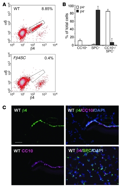
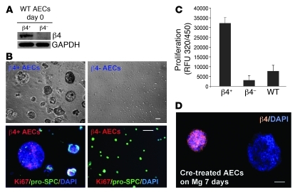
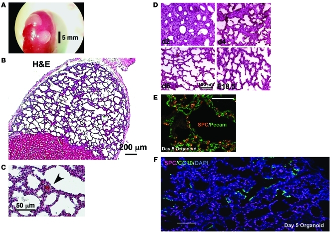
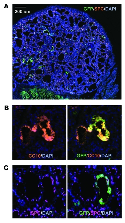
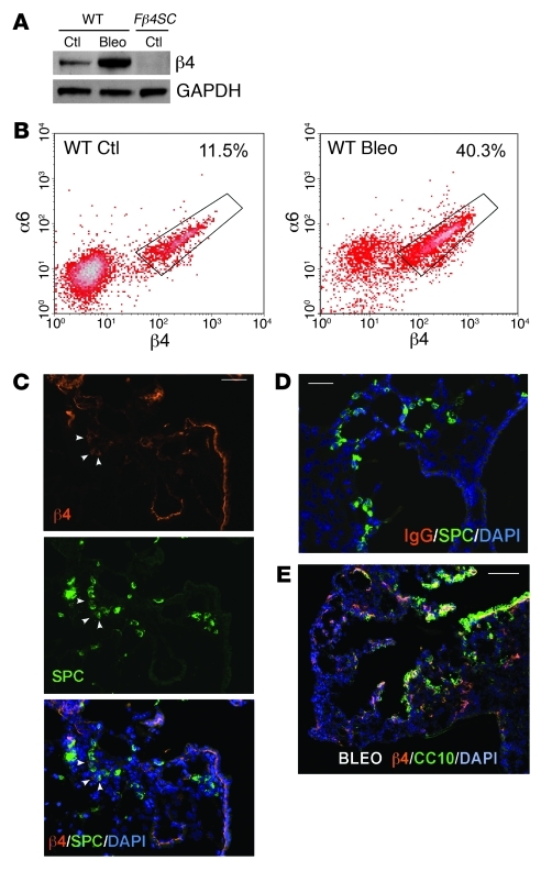
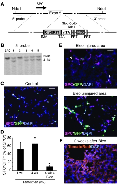
Laminins and their integrin receptors are implicated in epithelial cell differentiation and progenitor cell maintenance. We report here that a previously unrecognized subpopulation of mouse alveolar epithelial cells (AECs) expressing the laminin receptor α6β4, but little or no pro-surfactant C (pro-SPC), is endowed with regenerative potential. Ex vivo, this subpopulation expanded clonally as progenitors but also differentiated toward mature cell types. Integrin β4 itself was not required for AEC proliferation or differentiation. An in vivo embryonic lung organoid assay, which we believe to be novel, was used to show that purified β4+ adult AECs admixed with E14.5 lung single-cell suspensions and implanted under kidney capsules self-organized into distinct Clara cell 10-kDa secretory protein (CC10+) airway-like and SPC+ saccular structures within 6 days. Using a bleomycin model of lung injury and an SPC-driven inducible cre to fate-map AECs, we found the majority of type II AECs in fibrotic areas were not derived from preexisting type II AECs, demonstrating that SPC- progenitor cells replenished type II AECs during repair. Our findings support the idea that there is a stable AEC progenitor population in the adult lung, provide in vivo evidence of AEC progenitor cell differentiation after parenchymal injury, and identify a strong candidate progenitor cell for maintenance of type II AECs during lung repair.


Figures

Figure 1
Figure 1. Identification and location of β4+ AECs in murine lungs.
(A) FACS plot of α6 and β4 on AECs from WT and Fβ4SC mice. Residual β4 staining in the Fβ4SC mice is nonspecific secondary antibody. Percentages denote the fraction of cells within the boxed regions. (B) Differential cell counts of β4+ and β4 cells purified by flow cytometry from normal lungs (mean ± SD, n = 6). (C) β4 immunostaining in WT lungs showing diffusely stained cells near the bronchoalveolar junction along with CC10 staining and the merged images. Note that not all CC10+ cells were β4+. Basilar staining of airway β4 was apparent near the airway end of the junctional region. There was a lack of costaining of SPC with β4+ cells in the alveolar region (see also Supplemental Figure 3). Scale bar: 50 μm.
Figure 2
Figure 2. Ex vivo proliferation and clonal expansion of β4+ AECs.
(A) Immunoblot verifying separation of β4+ and β4 cells. (B) Phase-contrast images of β4+ and β4 AECs from WT mice cultured 7 days on Matrigel revealed large clusters only in the β4+ population. Ki67 and pro-SPC staining of cultured β4+ and β4 AECs revealed proliferation of β4+ cells and appearance of SPC+ cells, especially in the smaller clusters. Cells were initially SPClo or SPC. β4 AECs were strongly SPC+ and had little or no proliferation. The same number of β4+ and β4 cells were initially seeded. (C) BrdU assay at 24 hours (Chemicon) confirmed that greater than 90% of the proliferation among AECs resided in the β4+ fraction. WT, cell mixture before sorting. (D) AECs from floxed β4 mice treated ex vivo with adenovirus encoding cre recombinase. Developing clusters (>15 per culture well) were either all β4+ or all β4. Scale bars: 50 μm.
Figure 3
Figure 3. Embryonic lung organoid assay.
(A) Aggregates of single-cell preparations of E14.5 lung cells placed under renal capsules developed visible structures (white) after 5 days in vivo. The red structure is the underlying kidney. (B) Low-power view of H&E stains of the organoid revealed fluid-filled lacy structure under the capsule with visible airway-like and saccular elements. (C) Higher-power view revealed rbcs in blood vessel (arrowhead), suggestive of vascular continuity with the host kidney. (D) H&E-stained sections of day 2, day 4, and day 6 organoids showing development of the organoid to the saccular stage by day 6, similar to that of the E18.5 lung (see Supplemental Figure 5). (E) Distribution of SPC and Pecam-1 in a day 5 organoid. See Supplemental Figure 5 for distribution of Pecam-1 and SPC and absence of CC10 staining in E14.5 lungs. (F) CC10 and pro-SPC double staining of day 5 organoid sections. Airway-like regions led to SPC+ saccules with few, if any, SPC+CC10+ cells. Scale bars: 5 mm (A); 200 μm (B); 50 μm (C and E); 100 μm (D and F).
Figure 4
Figure 4. Development of adult β4+ AEC-derived structures in lung organoids.
(A) Low-power view of organoid with scattered clusters of GFP+β4+ AECs. Multiple images (original magnification, ×20) were captured and tiled into a single composite image. (B) Adjacent airway-like CC10+ structures, one completely GFP, the other entirely GFP+. Such dichotomy in the origin of CC10+ structures was the most common pattern in these mixed organoids. (C) SPC+ saccules, again showing entire saccular structures either GFP+ or GFP. Although this was the most common pattern, occasional saccules did contain both embryonic and adult SPC+ cells. Scale bars: 200 μm (A); 20 μm (B and C).
Figure 5
Figure 5. Expansion of β4+ AECs in lungs of bleomycin-treated mice.
(A) Immunoblot for β4 in purified β4+ AECs from mice injected with saline (Ctl) or bleomycin (Bleo) 10 days earlier. (B) The higher proportion of β4+ AECs from bleomycin-injected mice was confirmed by flow cytometry of β4 and α6. Percent cells within boxed regions is indicated. (C) β4 and SPC staining of a lung section from mouse injected intratracheally with bleomycin 14 days earlier. Arrowheads denote SPC+β4+ cells. (D) IgG-negative control for β4 immunostaining on a lung section adjacent to that in C. (E) Compared with normal lungs, there were many more β4+ cells in the alveolar compartment 21 days after bleomycin, but there were also CC10+β4+ cells extending into the alveolar region from the distal airway/bronchoalveolar junction. Scale bars: 50 mm (C and D); 100 μm (E).
Figure 6
Figure 6. Role for SPC progenitor cells in type II cell replenishment after bleomycin-induced injury.
(A) Insertion site of CreER2T, rtTA, and Neo cassette cDNAs into the stop codon of the endogenous SPC gene. A new stop codon was created at the 3′ end of rtTA, which was not used in these experiments. The Neo cassette had not been removed. See Supplemental Methods for details. (B) Southern blot of ES cell DNA using 5′ probe positioned as indicated in A revealed 4 correctly targeted clones (21-kb band in the Nde1 digest). (C) Merged image of pro-SPC and endogenous GFP fluorescence of SPCcreT2/loxp-tm-TR/GFP lungs 7 days after i.p. injection of 1 mg 4OH-tamoxifen. (D) Quantification of GFP+SPC+ cells as a fraction of total SPC+ cells in noninjured lungs (7 or 30 days after tamoxifen) or in injured regions of day 30 littermates injected with bleomycin 14 days before sacrifice. Greater than 200 SPC+ cells in more than 10 ×20 fields were counted in 3 sections per lung. Fibrotic areas were identified by crowded nuclei and distorted/collapsed alveolar architecture and confirmed by α-SMA staining of adjacent sections. *P < 0.0001. (E) Merged images of pro-SPC and GFP fluorescence of fibrotic and less-injured or uninjured lung areas of SPCcreT2/loxp-tm-TR/GFP mice 30 days after tamoxifen and 14 days after bleomycin. Arrowheads denote 2 GFP+SPC+ cells in fibrotic region. (F) Endogenous red fluorescence of bleomycin-injured loxp-tm-TR/GFP lung in the absence of cre. Scale bars: 50 μm.

Comment in

References

    1. Coraux C, Meneguzzi G, Rousselle P, Puchelle E, Gaillard D. Distribution of laminin 5, integrin receptors, and branching morphogenesis during human fetal lung development. Dev Dyn. 2002;225(2):176–185. - PubMed
    1. Nguyen NM, Senior RM. Laminin isoforms and lung development: all isoforms are not equal. Dev Biol. 2006;294(2):271–279. doi: 10.1016/j.ydbio.2006.03.032. - DOI - PubMed
    1. O’Reilly AM, Lee HH, Simon MA. Integrins control the positioning and proliferation of follicle stem cells in the Drosophila ovary. J Cell Biol. 2008;182(4):801–815. - PMC - PubMed
    1. Sheppard D. Epithelial integrins. Bioessays. 1996;18(8):655–660. - PubMed
    1. Wilhelmsen K, Litjens SH, Sonnenberg A. Multiple functions of the integrin alpha6beta4 in epidermal homeostasis and tumorigenesis. Mol Cell Biol. 2006;26(8):2877–2886. doi: 10.1128/MCB.26.8.2877-2886.2006. - DOI - PMC - PubMed

Publication types

MeSH terms