. 2011;33(3-4):299-311.
doi: 10.1159/000327243. Epub 2011 Jun 24.

Striatal neuroprotection from neonatal hypoxia-ischemia in piglets by antioxidant treatment with EUK-134 or edaravone

Affiliations

Striatal neuroprotection from neonatal hypoxia-ischemia in piglets by antioxidant treatment with EUK-134 or edaravone

Xinli Ni et al. Dev Neurosci. 2011.

Abstract

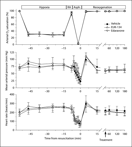
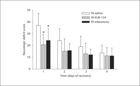
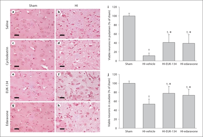
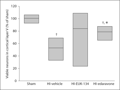
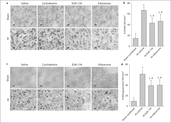
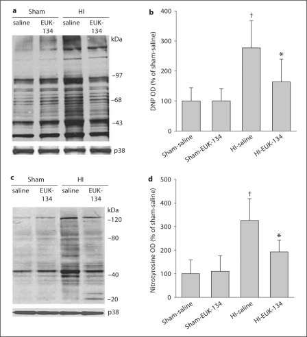
Striatal neurons are highly vulnerable to hypoxia-ischemia (HI) in term newborns. In a piglet model of HI, striatal neurons develop oxidative stress and organelle disruption by 3-6 h of recovery and ischemic cytopathology over 6-24 h of recovery. We tested the hypothesis that early treatment with the antioxidants EUK-134 (a manganese-salen derivative that acts as a scavenger of superoxide, hydrogen peroxide, nitric oxide or NO and peroxynitrite) or edaravone (MCI-186, a scavenger of hydroxyl radical and NO) protects striatal neurons from HI. Anesthetized newborn piglets were subjected to 40 min of hypoxia and 7 min of airway occlusion. At 30 min after resuscitation, the piglets received vehicle, EUK-134 or edaravone. Drug treatment did not affect arterial blood pressure, blood gases, blood glucose or rectal temperature. At 4 days of recovery, the density of viable neurons in the putamen of vehicle-treated piglets was 12 ± 6% (±SD) of sham-operated control density. Treatment with EUK-134 increased viability to 41 ± 17%, and treatment with edaravone increased viability to 39 ± 19%. In the caudate nucleus, neuronal viability was increased from 54 ± 11% in the vehicle group to 78 ± 15% in the EUK-134 group and to 73 ± 13% in the edaravone group. Antioxidant drug treatment accelerated recovery from neurologic deficits and decreased oxidative and nitrative damage to nucleic acids. Treatment with EUK-134 reduced the HI-induced formation of protein carbonyl groups and tyrosine nitration at 3 h of recovery. We conclude that systemic administration of antioxidant agents by 30 min after resuscitation from HI can reduce oxidative stress and salvage neurons in the highly vulnerable striatum in a large-animal model of neonatal HI. Therefore, oxidative stress is an important mechanism for this injury, and antioxidant therapy is a rational, mechanism-based approach to neuroprotection in the newborn brain.


Figures

Fig. 1
Fig. 1
Arterial O2 saturation, mean arterial pressure and heart rate are plotted for HI piglets treated with vehicle, EUK-134 or edaravone at 30 min of reoxygenation. Values are means ± SD measured: at baseline; at 5, 15, 27 and 37 min of hypoxia; at 5 min of room air (RA) ventilation; at each minute of a 7-min period of complete asphyxia (Asph.), and during 3 h of reoxygenation (note break and change in time scale).
Fig. 2
Fig. 2
Neurologic deficit scores (±SD) of piglets in the vehicle-treated, EUK-134-treated and edaravone-treated groups (n = 8 per group) at 1–4 days of recovery from HI. Maximum score = 154. * p < 0.05 vs. vehicle.
Fig. 3
Fig. 3
Neuropathology seen in HE-stained sections of piglet putamen 4 days after sham surgery or HI. In time-matched sham controls administered saline (a), 2-hydroxypropyl-β-cyclodextrin (c), EUK-134 (e) and edaravone (g), neurons appeared with a normal round shape, noncondensed nucleus and discrete nucleolus. The neuropil is smooth and uniform. In HI piglets treated with saline (b) and 2-hydroxypropyl-β-cyclodextrin (d), most of the principal striatal neurons underwent ischemic neurodegeneration, and the surrounding neuropil became pale and vacuolated. In HI piglets treated with EUK-134 (f) or edaravone (h), some of the neurons remained undamaged. Scale bars = 20 μm. Mean values ± SD of neuronal viability in putamen (i) and caudate nucleus (j) after sham surgery [combined cohorts with saline (n = 2), 2-hydroxypropyl-β-cyclodextrin (n = 2), EUK-134 (n = 2) and edaravone (n = 2)] or HI in piglets treated with vehicle [combined cohorts with saline (n = 4), 2-hydroxypropyl-β-cyclodextrin (n = 4)], EUK-134 (n = 8) or edaravone (n = 8). p < 0.05 vs. sham surgery; * p < 0.05 vs. HI-vehicle.
Fig. 4
Fig. 4
Density of viable neurons in layer V of primary sensorimotor cortex 4 days after sham surgery or HI in piglets treated with vehicle, EUK-134 or edaravone (n = 8 per group). Neuronal density was bimodal in the EUK-134 group, and the equal variance test failed. Data were analyzed by the Mann-Whitney nonparametric test, and results are displayed as a box plot (median and 25th and 75th percentiles). p < 0.05 vs. sham surgery; * p < 0.05 vs. HI-vehicle.
Fig. 5
Fig. 5
a In the sham groups, immunohistochemical analysis revealed faint staining for 8-OHdG within the cytoplasm and nucleus. The extent of staining for 8-OHdG was greater in the HI groups treated with saline or 2-hydroxypropyl-β-cyclodextrin vehicle than in the sham groups. Treatment of HI piglets with EUK-134 or edaravone reduced the staining. b Mean optical density (OD) values for 8-OHdG in the putamen of each group at 4 days of recovery. c Immunohistochemical analysis revealed no discrete positive staining for 8-nitroguanosine within the cytoplasm or nucleus in the sham groups. The extent of staining for 8-nitroguanosine was greater in the vehicle-treated HI groups than in the sham groups. Treatment of piglets with EUK-134 or edaravone reduced immunoreactivity after HI. d Mean OD values for 8-nitroguanosine in the putamen of each group at 4 days of recovery. a, c Scale bars = 10 μm. b, d Values are means ± SD (n = 8 per group). p < 0.05 vs. sham-combined; * p < 0.05 vs. HI-vehicle.
Fig. 6
Fig. 6
a Representative immunoblot showing DNP immunoreactivity as a measure of protein carbonylation in putamen sampled at 3 h of recovery from sham or HI piglets treated with saline or EUK-134. b Mean values ± SD (n = 4 per group) of total carbonyl optical density (OD) expressed as percentage of sham-saline values in putamen at 3 h of recovery. c Representative immunoblot of 3-nitrotyrosine immunoreactivity as a measure of protein nitration in putamen sampled at 3 h of recovery from sham or HI piglets treated with saline or EUK-134. d Mean values ± SD (n = 4 per group) of total nitrotyrosine OD expressed as percentage of sham-saline values in putamen at 3 h of recovery. a, c Synaptophysin (p38) was used as a protein loading control. b, d p < 0.05 vs. sham-saline, * p < 0.05 vs. HI-saline.

Similar articles

Cited by

References

    1. Ferriero DM. Neonatal brain injury. N Engl J Med. 2004;351:1985–1995. - PubMed
    1. Gonzalez FF, Ferriero DM. Therapeutics for neonatal brain injury. Pharmacol Ther. 2008;120:43–53. - PubMed
    1. Ferriero DM. Oxidant mechanisms in neonatal hypoxia-ischemia. Dev Neurosci. 2001;23:198–202. - PubMed
    1. Lafemina MJ, Sheldon RA, Ferriero DM. Acute hypoxia-ischemia results in hydrogen peroxide accumulation in neonatal but not adult mouse brain. Pediatr Res. 2006;59:680–683. - PubMed
    1. Sheldon RA, Christen S, Ferriero DM. Genetic and pharmacologic manipulation of oxidative stress after neonatal hypoxia-ischemia. Int J Dev Neurosci. 2008;26:87–92. - PMC - PubMed

Publication types

MeSH terms